Chapter 9 – synapses
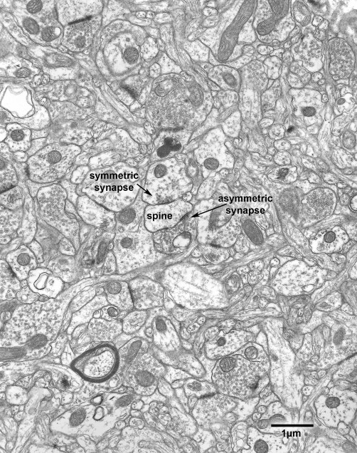
Basically, there are two types of synapses in the cerebral cortex, and in material fixed using glutaraldehyde they are referred to as symmetric and asymmetric synapses. The adjectives refer to whether the pre- and postsynaptic densities associated with the synaptic junctions are of similar width (symmetric), or whether the postsynaptic density is thicker (asymmetric) and more prominent than the presynaptic one (Figs. 9.1 -9.3).
Asymmetric synapses, which are excitatory in function, predominate and account for about 80% of the total population of synapses. Asymmetric synapses are formed by axon terminals that contain spherical synaptic vesicles. The synaptic junction has a rather wide cleft and an obvious postsynaptic density. The postsynaptic elements of such synapses include dendritic shafts, dendritic spines, and the cell bodies of inhibitory neurons.
The less common symmetric synapses are inhibitory in function. These synapses are formed by axon terminals that contain pleomorphic synaptic vesicles, which are somewhat smaller than the vesicles in axon terminals forming asymmetric synapses. At symmetric synapses the synaptic cleft is narrower than at excitatory synapses, and the postsynaptic density is only about the same width as the presynaptic density. The postsynaptic elements at such synapses include dendritic shafts (Fig. 9.2A and Fig. 9.4) some dendritic spines (Fig. 9.1 – 9.1B), the cell bodies of both excitatory and inhibitory neurons, and axon initial segments (Figs. 9.3).
It has been shown that with age some synapses are lost from the neuropil of prefrontal cortex area 46 and a few profiles of dystrophic axon terminals have been encountered in the cortical neuropil of old monkeys (Figs. 9.5 and 9.6). Generally the axoplasm of such terminals is filled with neurofilaments, sometimes surrounding a core of mitochondria (Fig. 9.6). That these profiles are of altered terminals is shown by the fact that they contain accumulations of synaptic vesicles at their peripheries, and are sometimes seen to be presynaptic to other structures in the neuropil. Whether there is an age-related loss of synapses from all cortical areas has not been determined. Other than area 46, the only other cortical structure in the monkey that appears to have been examined is the dentate gyrus, from which no age-related loss of synapses was found.
Figure 9.1
Neuropil in layer 5 of area 46 in a 30 year old monkey. In this field axon terminals are forming asymmetric and symmetric synapses with a dendritic spine.
Figure 9.1A
A version of Fig. 9.1 in which components of the neuropil have been colored. Axon terminals forming asymmetric synapses- light green; axon terminals forming symmetric, or inhibitory synapses- dark green; dendrites- blue; dendritic spines- grey; astrocytic processes- yellow. Profiles of axons have not been colored.
Figure 9.1B
An enlargement of part of Fig. 9.1. In the center of the micrograph is a dendritic spine (grey), which is synapsing with an axon terminal (light green) containing round synaptic vesicles that is forming an asymmetric synaptic junction. Another axon terminal (dark green) contains pleomorphic synaptic vesicles, and is forming a symmetric synaptic junction with the same dendritic spine. Other axon terminals in the fields are also forming asymmetric or excitatory synapses with dendritic shafts or spines.
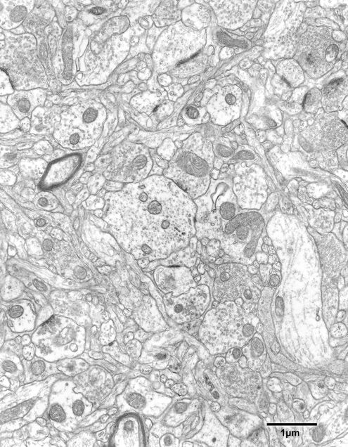
Figure 9.2
Neuropil in layer 5 of area 46 of a 25 year old monkey.
Figure 9.2A
A colorized version of Fig. 9.2. In the center of the field is an axon terminal (dark green) forming a symmetric, or inhibitory synapse with the shaft of a dendrite (blue). There is another axon terminal forming a symmetric synapse with a dendritic shaft in the lower right of the micrograph. All of the other axon terminals in the field (light green) contain round synaptic vesicles and are synapsing with either dendritic spines (grey) or with dendritic shafts (blue). As before, astrocytic processes are colored yellow and axonal processes have been left uncolored.
Figure 9.3
The axon initial segment of a pyramidal neuron in layer 4 of the visual cortex of a 6 year old monkey. The axon initial segment is forming symmetric, inhibitory synapses, with three axon terminals. These axon terminals are from a chandelier cell.
Figure 9.4
Neuropil in layer 2 of the primary visual cortex of a 25 year old monkey. In the middle of the field is a transversely sectioned dendritic shaft that is forming synapses with three axon terminals. One of the terminals contains pleomorphic synaptic vesicles and is forming a symmetric synapse with the dendrite. The other two axon terminals contain round, larger synaptic vesicles and are forming asymmetric synaptic junctions with the dendrite.
Figure 9.5
Neuropil in layer 6 of the primary visual cortex in a 25 year old monkey. In the middle of the field is the profile of a swollen axon terminal that appears to be degenerating. The profile is identified as belonging to an axon terminal by the presence of a cluster of synaptic vesicles in the cytoplasm on one side of the profile. On the other side of the profile is a row of mitochondria and the rest of the cytoplasm is filled with what appear to be neurofilaments.
Figure 9.6
A degenerating axon terminal in the neuropil of layer 2 in the primary visual cortex of a 35 year old monkey. The cytoplasm of the terminal has a core of clustered mitochondria surrounded by neurofilaments. At one side of the terminal is an aggregate of synaptic vesicles (arrows) where the terminal is forming asymmetric synapses with two dendritic spines.


