Chapter 6 – myelin sheaths with dense degeneration
Age-related structural alterations in myelin sheaths are of two types. The most common form of alteration is splitting of the myelin lamellae at the major dense line to accommodate pockets of dense cytoplasm (Fig. 6.1), and since these splits occur at the major dense line, the electron dense cytoplasm within the splits must be derived from the oligodendrocyte forming the sheath. This type of myelin degeneration is referred to as dense degeneration.
In dense degeneration the extent of the splitting is very variable. In the mildest cases the split is not very extensive and occurs between one or two adjacent myelin lamellae (Fig. 6.1), but even when a single split occurs there can be extensive accumulation of dense cytoplasm in the split causing the sheath to bulge out (Fig. 6.2 and 6.2A). But in more extreme cases the splitting affects a number of lamellae (Figs. 6.3 and 6.4). Sometimes vacuoles occur in the dense cytoplasm (Fig. 6.5), and in other examples the extensive dense cytoplasm contains organelles that appear to be a mixture of vacuoles and lysosomes (Figs. 6.6 , 6.6A and 6.7). In all of these examples the axon is still intact, even though it may be pushed to one side of the enlarged nerve fiber profile.
Longitudinal sections show that these splits of sheaths that contain dense cytoplasm are localized, so that they produce bulges on the outsides of the myelin sheaths (Figs. 6.8 and 6.9). In some examples, several pockets of dense cytoplasm are evident along the same internodal length of myelin (Fig. 6.10).
Figure 6.1
A split myelin sheath surrounding an axon in layer 4 of area 46 in prefrontal cortex of a 35 year old monkey. The split has occurred along the major dense line of the sheath, and it contains some dense cytoplasm that must belong to the parent oligodendrocyte.

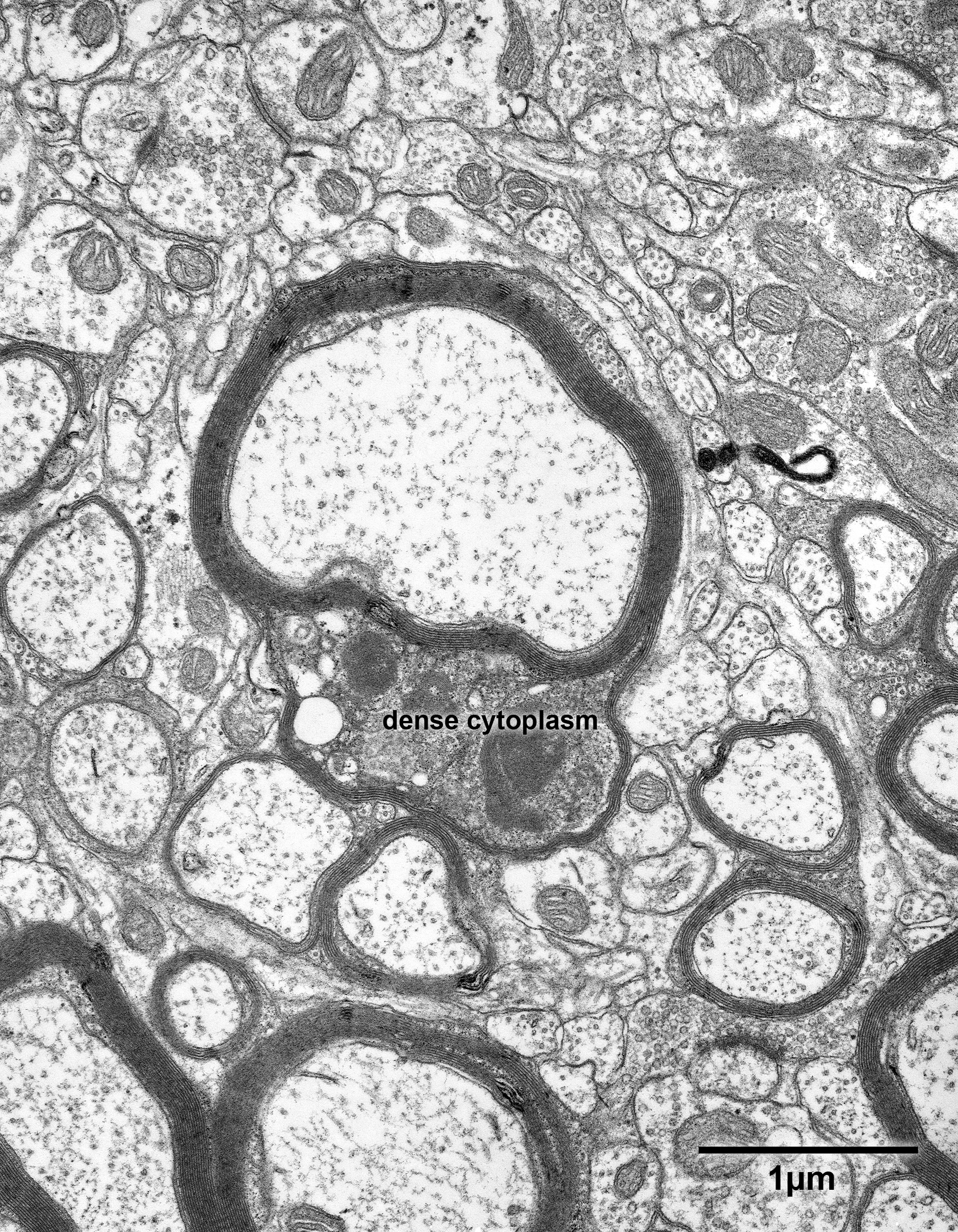
Figure 6.2
A split myelin sheath in layer 4 of the primary visual cortex of a 35 year old monkey. The split is localized and produces a bulging of the sheath. The dense cytoplasm in the split is granular and contains vacuoles and lysosomes.

Figure 6.2A
A copy of Fig. 6.2 in which some of the components have been colored.
Astrocytes – yellow; unmyelinated axons- light green; axon terminals- dark green; oligodendrocytic processes- red: dendrites- blue; dendritic spines- grey.

Figure 6.3
A bundle of nerve fibers in layer 4 of the primary visual cortex of a 30 year old monkey. One of the axons (axon) is surrounded by a sheath that has a large and localized bulge that contains dense cytoplasm. In this example the bulge is produced by the splitting of several lamellae.

Figure 6.4
A bundle of myelinated nerve fibers in layer 4 of the primary visual cortex of a 30 year old monkey. While most of the nerve fibers are normal a few show age changes. Three of the nerve fibers show splitting of the lamellae with accumulation of dense cytoplasm in the splits, while two other nerve fibers have sheaths with redundant myelin.

Figure 6.5
Nerve fibers in layer 4 of area 46 of a 35 year old monkey. One of the nerve fibers has a sheath with a split that contains dense cytoplasm with two vacuoles.

Figure 6.6
A myelinated nerve fiber in layer 4 of area 46 from a 24 year old monkey. On one side of the nerve fiber the myelin sheath shows extensive splitting between several myelin lamellae, with a massive accumulation of dense cytoplasm.

Fig. 6.6A
A colored version of 6.6. Oligodendrocytes- red; myelinated axons- dark green; dendrites- blue; dendritic spines- grey; axon terminals- pale green.

Figure 6.7
A large myelinated nerve fiber in layer 4 of area 46 in a 25 year old monkey. The axon appears to be the structure in the lower part of the profile. The space between the axon and the myelin sheath contains a variety of unidentifiable components embedded in a granular, dark cytoplasm.

Figure 6.8
A longitudinally sectioned myelinated nerve fiber in layer 4 for area 46 in a 35 year old monkey. It is apparent that accumulations of dense cytoplasm and myelin balloons are localized within small areas of the myelin sheaths, and that both degenerative changes can be present side-by-side.

Figure 6.9
A longitudinally sectioned myelinated nerve fiber in layer 4 of area 46 from a 30 year old monkey. The nerve fiber shows a large and localized accumulation of dense cytoplasm in a split of the myelin sheath.

Figure 6.10
A longitudinally sectioned myelinated nerve fiber in layer 4C of the primary visual cortex of 35 year old monkey. The nerve fiber has two locations, on the right of the image, in which the myelin sheath is split to accommodate dense cytoplasm.
