Chapter 14 – microglial cells
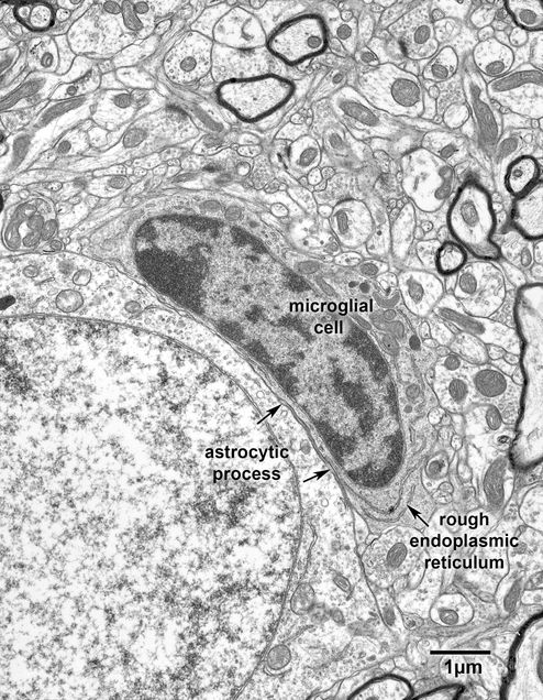
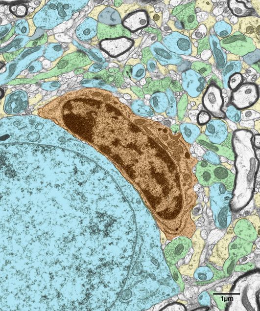
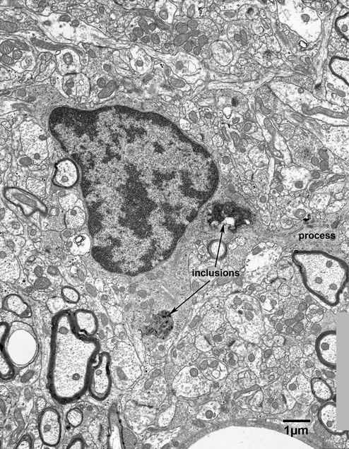
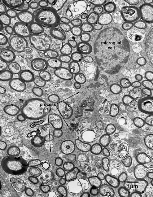
Like oligodendrocytes, microglial cells have dark nuclei with clumped chromatin and dark cytoplasm. However, there are subtle morphological differences between these two neuroglial cell types. The nuclei of microglial cells in young monkeys tend to be more elongated, the contours of the cell bodies are more irregular than those of oligodendrocytes and although not always apparent (Fig. 14.1) the cisternae of the rough endoplasmic reticulum in microglial cells are much longer than those of oligodendrocytes (Figs. 14.2 and 14.2A). Furthermore when oligodendrocytes are adjacent to neurons the plasma membranes of the two cells are closely apposed, whereas when microglial cells lie adjacent to neurons it is usual to find a thin astrocytic processes intervening between the two cells (Figs 14.1 and 14.2A). Sometimes processes can be seen to extend from microglial cells (Fig. 14.3 and 14.4). These tend to have irregular contours and extend branches into the surrounding neuropil.
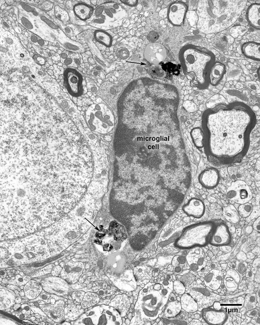
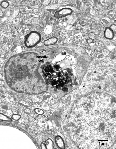
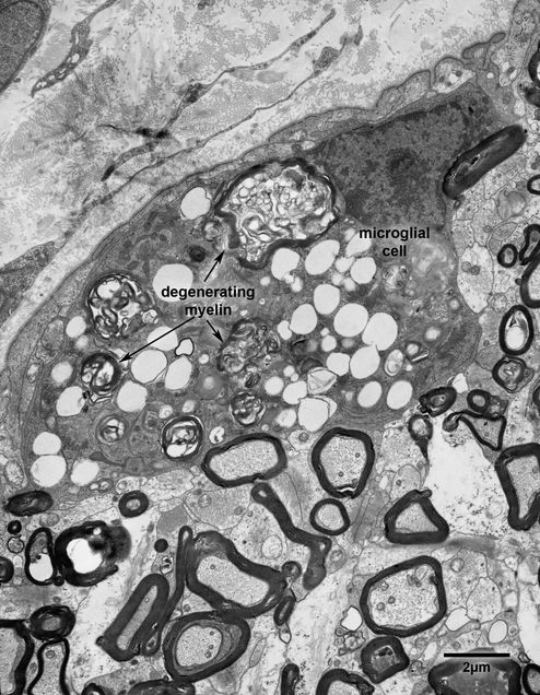
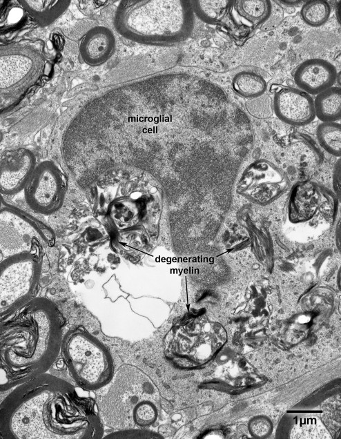
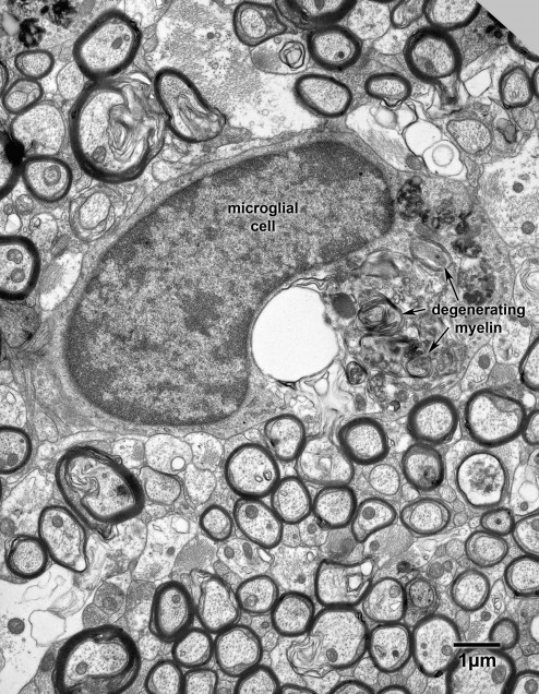
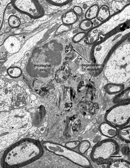
Microglial cells are phagocytes and so in older monkeys it is common to find inclusions in their perikarya and processes. In cerebral cortex the inclusions tend to be simple in form and resemble lipofuscin (Figs. 14.5), although they may be voluminous (Figs. 14.6 and 14.6A), whereas in optic nerves (Fig. 14.7) and corpus callosum (Fig. 14.8), and fornix (Fig. 14.9) of old monkeys, it is evident that some of the inclusions are derived from the phagocytosis of myelin. In yet other parts of the nervous system, such as the spinal cord (Fig. 14.10), the appearance of the inclusions is quite different, although they still seem to be derived from degenerating myelin.
Figure 14.1
A microglial cell adjacent to a neuron on layer 4 of the primary visual cortex of a 15 year old monkey. Although microglial cells are dark like oligodendrocytes, they usually have bean shaped nuclei and the contours of their cell bodies are more irregular than those of oligodendrocytes.
Figure 14.2
A microglial cell adjacent to a neuron in the primary visual cortex of a 5 year old monkey. This micrograph shows two important features of microglial cells, namely the long cisternae of rough endoplasmic reticulum and the fact when a microglial cell is adjacent to a neuron there is usually a thin astrocytic process (arrow) between the two cells.
Figure 14.2A
A version of Fig. 14.2 in which some of the elements have been colored. Microglial cell- orange; dendrites- blue; axon terminals- green; dendritic spines- grey.
Figure 14.3
A microglial cell in the primary visual cortex of a 35 year old monkey. This micrograph shows the irregular contours of a typical microglial cell and the features of the processes that emanate from these cells. Note the presence of the dark amorphous inclusions in the perikaryal cytoplasm.
Figure 14.4
A microglial cell in the fornix of a 32 year old monkey. Note the branching process extending from the lower border of the cell (arrow) and the presence of numerous inclusions in the cytoplasm of the process.
Figure 14.5
A microglial cell in the primary visual cortex of a 35 year old monkey. Note the phagocytozed material at the two poles of the perikaryon (arrows).
Figure 14.6
A microglial cell in layer 3 of the primary visual cortex of a 25 year old monkey. The perikaryon of this cell is distended by a large inclusion of phagocytozed material.
Figure 14.6A
A colored version of the electron micrograph shown in Fig. 14.6. Microglial cell- orange; neuronal cell body and dendrites- blue; axon terminal- green; dendritic spines- grey; astrocytic processes- yellow.
Figure 14.7
A microglial cell in the optic nerve of a 32 year old monkey. The microglial cell has phagocytozed a large amount of debris, some of which is obviously degenerated myelin.
Figure 14.8
A phagocytic microglial cell in the corpus callosum of a 35 year old monkey. The perikaryon is full of phagocytozed material, some of which appears to be degenerating myelin.
Figure 14.9
A phagocytic microglial cell in the fornix of a 32 year old monkey. Again, much of the phagocytozed material appears to be degenerating myelin.
Figure 14.10
A phagocytic microglial cell in the spinal cord of a 35 year old monkey. Many of the inclusions have the appearance of degenerating myelin.


