Histomorphologic Analysis of Ebola Viral Disease in Rhesus Macaques Using Deep Learning
SPRING 2020 RESEARCH INCUBATION AWARDEES
PI: Nicholas Crossland, Assistant Professor, Pathology and Laboratory Medicine, MED
Co-PI: Vijaya Kolachalama, Assistant Professor, Medicine, MED
Track: Hariri Institute for Computing
What is the Challenge?
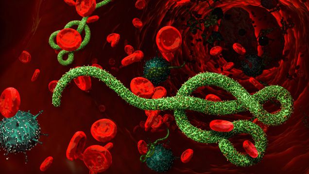
Ebola viral disease (EVD) is caused by 1 of 4 species in the Ebolavirus genus (Zaire, Sudan, Taï Forest, & Bundibugyo). These result in hemorrhagic fever in humans. The fatality rate ranges from 25-90%. In the West African epidemic that took place from 2014-2016, 28,652 cases and 11,325 deaths occurred in Guinea, Liberia, and Sierra Leone, with an estimated $53 billion cost to the global economy. The second-largest EVD outbreak ever recorded is currently being resolved in the Democratic Republic of Congo. This includes 3,453 cases and 2,264 deaths as of April 5th, 2020. Notably, this was the first outbreak where a highly effective and safe vaccine was distributed with over 223,000 people who have received the vaccine during the outbreak. Unfortunately, the vaccine is only efficacious against EBOV and not the other three species of Ebolavirus. Thus EVD remains a significant emerging public health threat.

What is the Solution?
Nicholas Crossland, along with Co-PI, Vijaya Kolachalama, a Research and Junior Faculty Fellow at the Hariri Institute of Computing aim to generate new analytical tools for understanding biological data sets and the interrelatedness or quantitative histomorphology “pathomics” with other clinicopathological study data. Furthermore, their research represents the opportunity to catalyze the collaboration between two faculty members with unique but synergistic areas of expertise, pathology, and the computer sciences. This collaboration would lead to the ability to autonomously classify and quantify EVD-associated histomorphological phenotypes.

What is the Process?
The team would use a combination of longitudinal samples collected from the liver and spleen of rhesus macaques, gathered over 3-8 days post-infection (DPI), in addition to terminal animals (6-8 DPI) that were euthanized due to clinical deterioration. These samples have been chemically inactivated and transported to Dr. Crossland’s BSL-2 laboratory and are available immediately for Model Table 1. Ebola Viral Disease Histomorphologic Phenotypes. It is proposed that Histomorphological phenotypes of EVD would autonomously classify and quantify with the use of deep learning.