Recent ENG PhD Helps Develop Highly Sensitive Imaging Technique to Detect Myelin Damage
The breakdown of myelin, the insulating layer around brain cells that supports brain function, is prevalent in a range of neurodegenerative diseases, aging and because of various forms of trauma. While electron microscopy is considered the gold standard for ultrastructural imaging of myelin, it is considered impractical for large-scale studies due to its limited field of view and time-consuming and complex sample preparation requirements.
In a new study from Boston University Chobanian & Avedisian School of Medicine and BU’s College of Engineering, researchers used a special microscope called birefringence microscopy (BRM) paired with an automated deep learning algorithm to reliably count and map myelin damage across whole sections of the brain—something not feasible with other techniques. The ability to image and measure damage to myelin will lead to better understanding the patterns and extent that occurs with disease, injury and normal aging.

“A major advantage of BRM over conventional imaging methods is its ability to rapidly image large areas at high resolution without special staining, making it uniquely suited for studying widespread myelin pathology,” says corresponding author Alex Gray, who recevied his PhD in BME in 2025 and was advised by Professor Irving Bigio.
Gray is currently employed at Analog Devices | Analog Garage (an innovation hub within Analog Devices) as a research scientist/engineer, working on exploring novel technologies in the healthcare space.
“I started at BU as a masters student in Irving Bigio’s lab interested in biomedical optics for healthcare applications. I worked on a miniaturized elastic scattering spectroscopy device intended to improve surgical workflows for assessing cancer margins during oral cancer surgical resection,” Gray said. “I owe a lot to my mentors and training at BU where I learned how to approach complex problems holistically, which is a requirement in the healthcare space.”
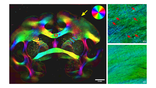 Gray and the team developed a technique that combines birefringence microscopy (BRM), a method to image myelin in brain slices, and deep learning to study myelin damage in different models of neurodegeneration – where myelin damage is thought to be related to functional deficits like cognitive impairment or motor function loss. In a model of stroke-like brain injury, this method allowed them to rapidly scan large areas of brain tissue and automatically measure myelin damage without using complex stains. This technique demonstrated that treatment with stem-cell derived vesicles reduced myelin damage (or promoted repair), a task that would be much more difficult or impractical with other imaging techniques, demonstrating how this tool can help researchers track the mechanisms of myelin damage in different neurodegenerative disease and test therapies for conditions like stroke, Alzheimer’s, and multiple sclerosis.
Read the article in Neurophotonics