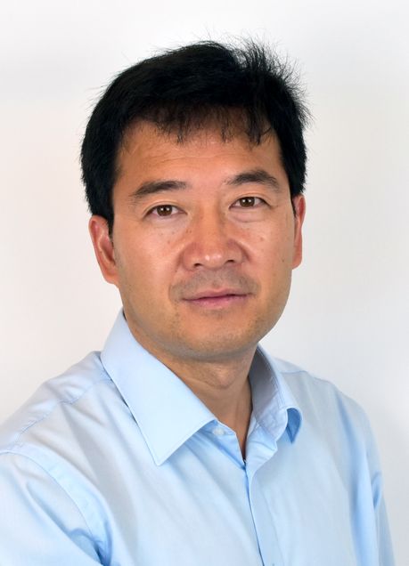
Cheng Wins Grant to Continue Breakthrough Imaging Research
By Liz Sheeley

Professor Ji-Xin Cheng (ECE, BME, MSE) wants to make diagnostic imaging better by following the biology. He sees his work to create imaging systems that don’t rely on biological markers as a way to answer questions we don’t even have yet, using label-free imaging. This technique allows clinicians to see a bigger picture, and not just what they’re looking for.
This summer, Cheng received a Maximizing Investigator’s Research Award (MIRA) for $2.9 million over five years from the National Institutes of Health (NIH) to continue his work. Investigators receive MIRAs after a successful use of the NIH’s top research grant, the R01—the MIRA is a type of extension on that grant that allows researchers to build on their work.
To receive this grant, the NIH looks at what an investigator has done in the past five years. For Cheng, he’s devoted that time to inventing and advancing two novel imaging techniques, making them better by increasing precision and specificity all while figuring out how to get them into the clinic.
These techniques – multiplex simulated Raman scattering (SRS) microscopy and mid-infrared photothermal (MIP) microscopy – can map cell metabolism to better understand the development of cancer and many other diseases, help physicians accurately remove cancerous cells in surgery and quickly test for antibiotic effectiveness.
And because they are label-free – a technique used when the researcher or physician doesn’t know what the target of the imaging should be — they can apply to a wide range of cancers and diseases. For example, when a physician is looking to diagnose a patient and suspects cancer, there are specific biological markers to confirm a diagnosis. Cancer is not just one disease and scientists don’t definitively know which tests to run in order to be sure that a patient is cancer free. Label-free imaging takes that uncertainty away by being able to image a patient’s cells to look for signs of cancer, like increased metabolic rate, without requiring a specific molecular target.
His laboratory is working on bringing SRS imaging into the clinic by developing a handheld SRS microscope. The new tool was able to detect the difference between cancerous and non-cancerous brain tissue. Infrared photothermal microscopy is a technique Cheng is developing that can map the metabolism of cells to potentially aid in disease diagnoses and treatment.
In his early career, Cheng has spearheaded the development of coherent anti-Stokes Raman scattering (CARS) microscopy and demonstrated label-free CARS imaging of the myelin sheath, the insulating layer that wraps around nerve fibers, to look at spinal cord injuries, and triggered the development of a nanomedicine for functional recovery of an injured spinal cord.
When Cheng was a postdoc at Harvard University in the department of chemistry and chemical biology, he sat in undergraduate classes in biology and physiology to help him understand the gaps in biologists’ techniques and tools. Previously, he had studied physics and chemistry during his undergraduate and graduate studies.
“My advisor, Professor Xiaoliang Sunney Xie, always said you could develop a tool, but it will not be useful unless biologists use it,” says Cheng. “There are large breakthroughs in biology that were serendipitous and not driven by design, so by expanding the ability of scientists to look beyond what they know, we can allow for more of those discoveries to happen.”