Brain Systems for Language
![]()
The goal of this study was to understand the range of language abilities and brain systems that may explain why people with Autism Spectrum Disorders have difficulties communicating with spoken language.
How the brain processes sound
Everyday listening is a very demanding activity! We need to separate out all the different sounds in the environment in order to hear the speech of someone talking to us. This involves complex brain processing that we can monitor by investigating electrical activity in the brain. In this study we measure activity using ‘electroencephalography’ – or EEG collected through small soft probes embedded in a specially designed, comfortable cap. Participants will hear different sounds playing in the room while we record brain activity.
How the brain produces speech

Speaking seems so effortless that we don’t realize what a complex process it is! Many brain areas are involved in this process, and one important key to producing speech is the integration of activity across these brain areas. In this study we use magnetic resonance imaging (MRI) to investigate the connections in the brain that are related to producing speech. We are especially interested to see whether there are specific problems in the connections between the brain areas involved in initiating speech. Participants will lie in the scanner, which is located at MGH, and be asked to name pictures or to just relax if they have no spoken language.
Who participated?
Children, adolescents and young adults with ASD between the ages of 8 – 21 and adolescent and young adults without ASD between the ages of 8 and 21.
What did participants do?
Participation involved multiple visits to BU and MGH. We worked with families to create a schedule that fit their needs and preferences. Enrolled families completed thorough pre-visit screens and the following:
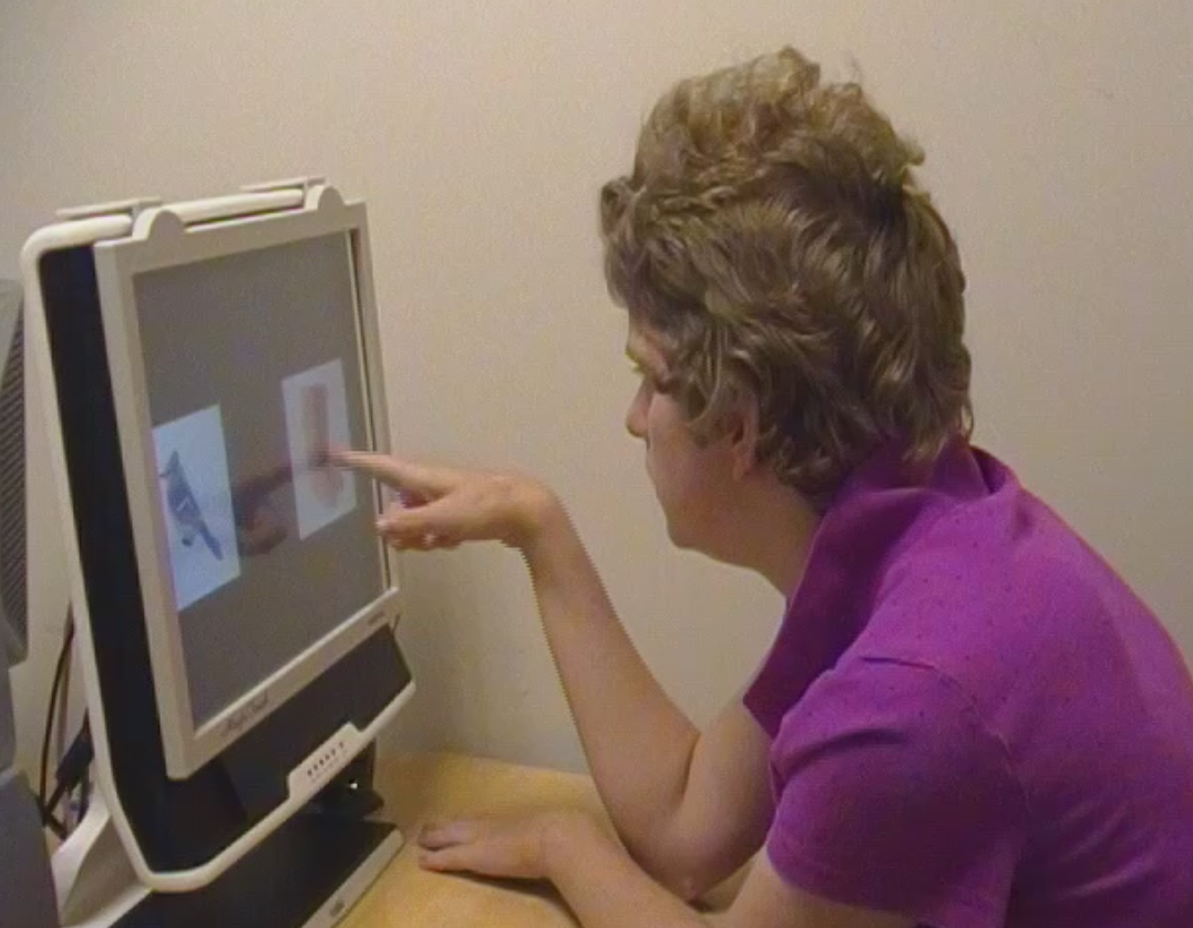 Behavioral Testing
Complete a battery of standardized language and cognitive assessments.

EEG Testing
Participants watched a video of their choice on mute while listening passively to different sounds.
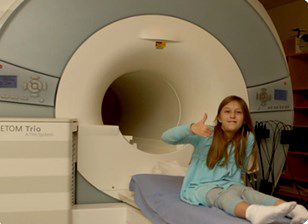
Brain Imaging
Participants worked with staff members using play materials to simulate the scanner space and sounds at BU. Once participants were comfortable and wiggle-free in this setting, we scheduled a visit to MGH to practice in a mock scanner. This looks just like to real scanner but has no magnet. Eventually we scheduled the real MRI at MGH!
Where and when?
Sessions were held at the Center for Autism Research Excellence at BU in Boston, and at the Martinos Brain Imaging Center in the Charlestown Navy Yard.