Tim Weber Successfully Defends his PhD
Congratulations to Tim Weber for successfully defending his PhD on “Transillumination Techniques in Ophthalmic Imaging” !
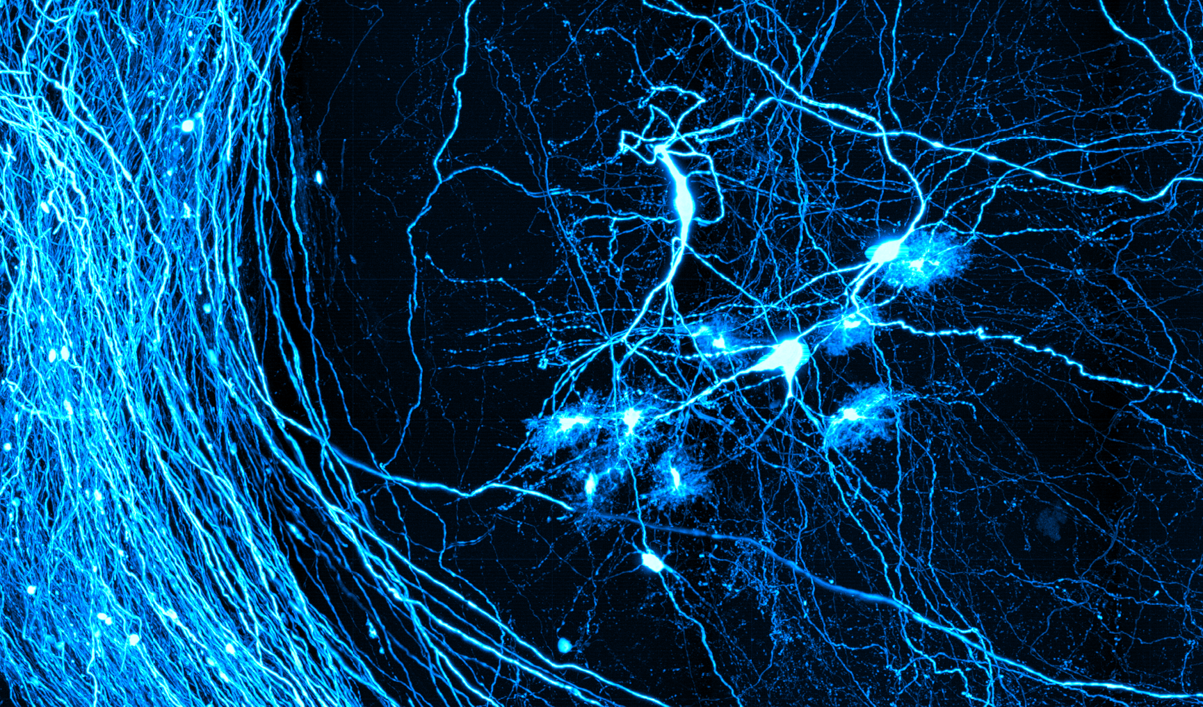
Tim Weber (Mertz lab) is an engineer by training and focuses on developing new microscopes and imaging methods for the life sciences. He received his B.S. in Biomedical Engineering and Applied Mathematics from Columbia University in 2014. He was drawn to Neurophotonics during the development of Active Illumination Microscopy, a method for greatly boosting the dynamic range of a scanning microscope. The technique has proved extremely useful for two-photon imaging of bright neural cell bodies and faint dendrites in the same field of view. Currently, he labors away at his dissertation on switched multi-region microscopy and a separate new imaging method for ophthalmology. He works for Jerome Mertz in the Biomicroscopy Lab
Dissertation Abstract:
In vivo imaging of the human cornea and retina is typically performed in a reflection geometry. Images are formed from light that has backscattered off corneal microstructures or backreflected from the retina. In this configuration, artifacts caused by superficial surface reflections are often encountered. These unwanted reflections can either globally overwhelm the signal or cause local glare, complicating reliable image quantification. This thesis describes a pair of alternative ophthalmic imaging techniques based instead on transmitted light, which inherently avoids these artifacts.
For retinal (i.e. fundus) imaging, we describe a mesoscopic transmission imaging method, which we call transcranial fundus imaging. The method uses deeply penetrating near-infrared light delivered transcranially from the side of the head, and exploits multiple scattering to redirect a portion of the light towards the posterior eye. This unique transmission geometry simplifies absorption measurements and enables flash-free, non-mydriatic imaging as deep as the choroid. We use multispectral image sets taken with this new transillumination approach to estimate oxygen saturation in retinal blood vessels.
In the cornea, we describe a new technique for non-contact phase-contrast microscopic imaging. It is based on fundus retro-reflection and back-illumination of the crystalline lens and cornea. To enhance phase-gradient contrast, we apply asymmetric illumination by illuminating one side of the fundus. The technique produces micron-scale lateral resolution across a 1-mm diagonal field of view. We show representative images of the epithelium, the subbasal nerve plexus, large stromal nerves, dendritic immune cells, endothelial nuclei, and the anterior crystalline lens, demonstrating the potential of this instrument for clinical applications.