TRD #3 – Anna Devor – Neurophotonic devices for microscopic neurovascular measurements
Specific Aims
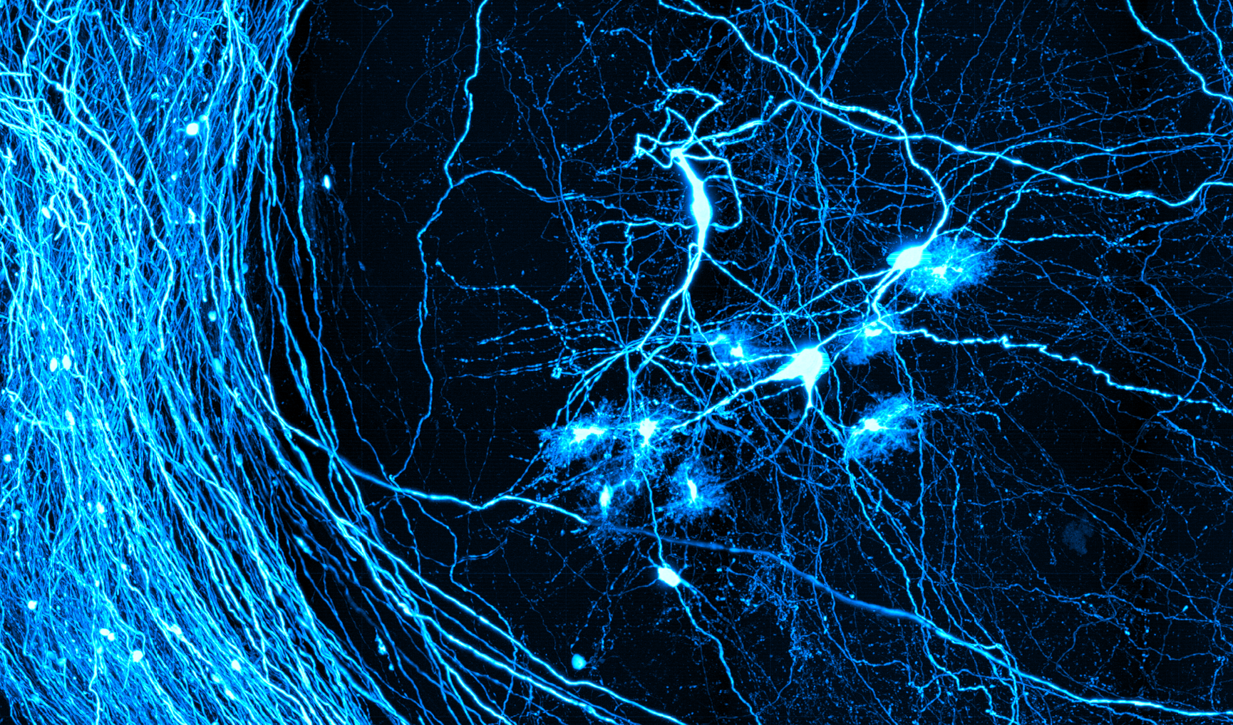
During the last two decades, we have been at the forefront of optical microscopy developments that have enabled a versatile suite of tools for high-resolution, high-sensitivity measurements of neurovascular and neurometabolic parameters in cerebral cortex of experimental animals in vivo. Our ongoing efforts include combining optical and electrical sensing, increasing the accuracy of quantification of cerebral blood flow across different types of blood vessels, and enabling quantification of O2 consumption. Collaborative projects are driving the need for further advancement of these technologies to improve the spatial coverage and depth penetration while maintaining or increasing temporal resolution and improving reliability of the measurements in the context of longitudinal imaging in awake behaving mice.
Working in close push-pull collaboration with our CPs, we will advance our methods for microscopic and mesoscopic measurements neuronal (Aim 1), metabolic (Aim 2), and vascular (Aim 3) activity associated with normal function and pathology of the neurovascular unit (NVU) to impact studies of neurovascular coupling in health and in disease. Methods include multiplexed electrophysiological recordings with transparent electrode arrays, deep multiphoton microscopy for O2 imaging, and mesoscopic multimodal measurements of neuronal activity and blood flow. Taken together, these aims bear on the key physiological parameters underlying noninvasive imaging signals of TRD #1 and TRD #2. The aims are complementary in that the different measurement modalities can be combined to permit comprehensive probing of the NVU activity in health and disease while also providing foundation for interpretation of measurements delivered by TRD #1 and TRD #2.
Aim 1: Scale up our wearable, transparent electrode array and deploy this technology for chronic multimodal recordings combined with optical imaging. The goal is to enable robust multimodal, large-scale investigation of neurovascular and neurometabolic dynamics through seamless integration of electrode array recordings with single- and multiphoton optical imaging methods.
- Extend coverage to the entire dorsal surface of the mouse brain (up to 128 contacts per hemisphere) acting as artificial and optically transparent dura.
- Engineer one or more laminar (penetrating) electrodes per hemisphere while optimizing for optical clarity. Each laminar electrode will be composed of 10 contacts spaced by 100 μm along a penetrating 1-mm shank, integrated with surface arrays in a single device.
Aim 2: Push the depth penetration of 2-photon phosphorescence lifetime microscopy (2PLM) for quantification of tissue partial pressure of O2 (pO2) and combine 2PLM with fluorescence imaging. The goal is to enablequantification of pO2 and O2 metabolism throughout the cortical depth in the context of vasodilation and brain state dynamics reflected by specific fluorescent probes.
- Increase depth penetration of 2PLM by implementing non-degenerate 2-photon (ND2P) excitation with 2 laser beams of different color, which will be sculpted to overlap in space only at the focal plane.
- Enable rapid transition between different scanning modes to interleave point-scan 2PLM with line- or frame-scan 2-photon fluorescence imaging.
Aim 3: Advance our new Dynamic Laser Speckle Imaging (DLSI) method for quantitative high-speed 2D measurements of blood flow with mesoscopic resolution to provide axially resolved 3D measurements of blood flow dynamics using a novel interferometric extension of the technology. This Aim will also deliver a new interference speckle spatial statistics analysis for quantification of the speckle decorrelation time that will be leveraged by TRD #2.
- Develop novel interferometric DLSI (iDLSI) and interferometric Laser Speckle Contrast Imaging (iLSCI) capable of acquiring over 2048x2048x50 blood flow voxels/s. These methods will be up to 700 times faster compared to conventional point scanning spectral domain optical coherence tomography (OCT).
- Integrate iDLSI/iLSCI with mesoscopic 1-photon fluorescence imaging and imaging of hemoglobin absorption to be disseminated within a single instrument.
For each one of the Aims, we will be working in close collaboration with our CPs to optimize and stabilize these technologies. We will deliver devices, protocols, procedures, tutorial, software, and host researchers to aid our SPsand the broader community in establishing the technology in their own labs. In parallel, our deliverables will help interpretation of noninvasive imaging being advanced by TRD #1&2.