How Ultrafast Lasers Developed in Michelle Sander’s Lab are Speeding Up Biological Research | Faculty Spotlight
By Kat J. McAlpine, Photos by Christopher McIntosh
Michelle Sander and her lab group at Boston University are seeking out new ways to build ultrafast lasers – lasers that emit such short-lasting pulses of light that their duration is difficult to fathom.
“I’m particularly interested in developing lasers on the femtosecond scale – a fraction of a second so small that its ratio is the same as one second to 32 million years,” Sander says.
 At these incredibly ultra-short timescales, Sander and her team are interested in the way light and matter interact. “When you shine light onto a material, you can characterize that material or change its properties,” she says. “Light pulses can travel up to 300 nanometers within one femtosecond – a distance that’s just a fraction of the width of a human hair – and create new phenomena that we don’t see with longer-lasting or continuous laser light. We’re working to design novel lasers that can emit these femtosecond pulses and to better understand the formation of these pulses and their effect on materials.”
Sander, who at BU is a faculty member at the Photonics and Neurophotonics Centers and a College of Engineering associate professor of electrical and computer engineering, launched her lab more than 10 years ago after earning a PhD from Massachusetts Institute of Technology. But her career in engineering began at an early age. When she was a young student growing up in Germany, she remembers being fascinated by physics and technology during high school.
“There was a program aimed at recruiting more women in STEM fields, offering hands-on experiences in different types of labs,” she says. “At that time, less than 5 percent of electrical engineering students were women.” Sander jumped at the opportunity to intern in an electrical engineering lab at a local university, which helped cement her interest in the field. After high school, she started taking university-level engineering courses in Germany, and then earned a Fulbright fellowship that brought her to the Georgia Institute of Technology in Atlanta for graduate studies.
Today, at BU, Sander’s team is looking beyond fundamental research to develop practical, real-world applications based on various lasers and further elucidate what these lasers can reveal about different materials. She considers her team to be a “research family,” made up of many deep connections within the group that have developed through intense discussions about science and common goals.
“I think it’s really important to have an atmosphere where people can get direct feedback and also communicate directly with others,” says Sander, who is also affiliated with BU’s biomedical engineering and materials science and engineering departments. “Every paper a lab member publishes, every presentation a student gives at a conference, these are really exciting and meaningful moments for our team.”
She invites innovation and creativity into the lab and says it’s hard to predict which direction the team’s research will go in – she and her team are always open to new ideas and external partnerships.
When optics, physics, and biology collide
Right now, the team is excited about several multi-institutional collaborations that apply their expertise towards biological applications.
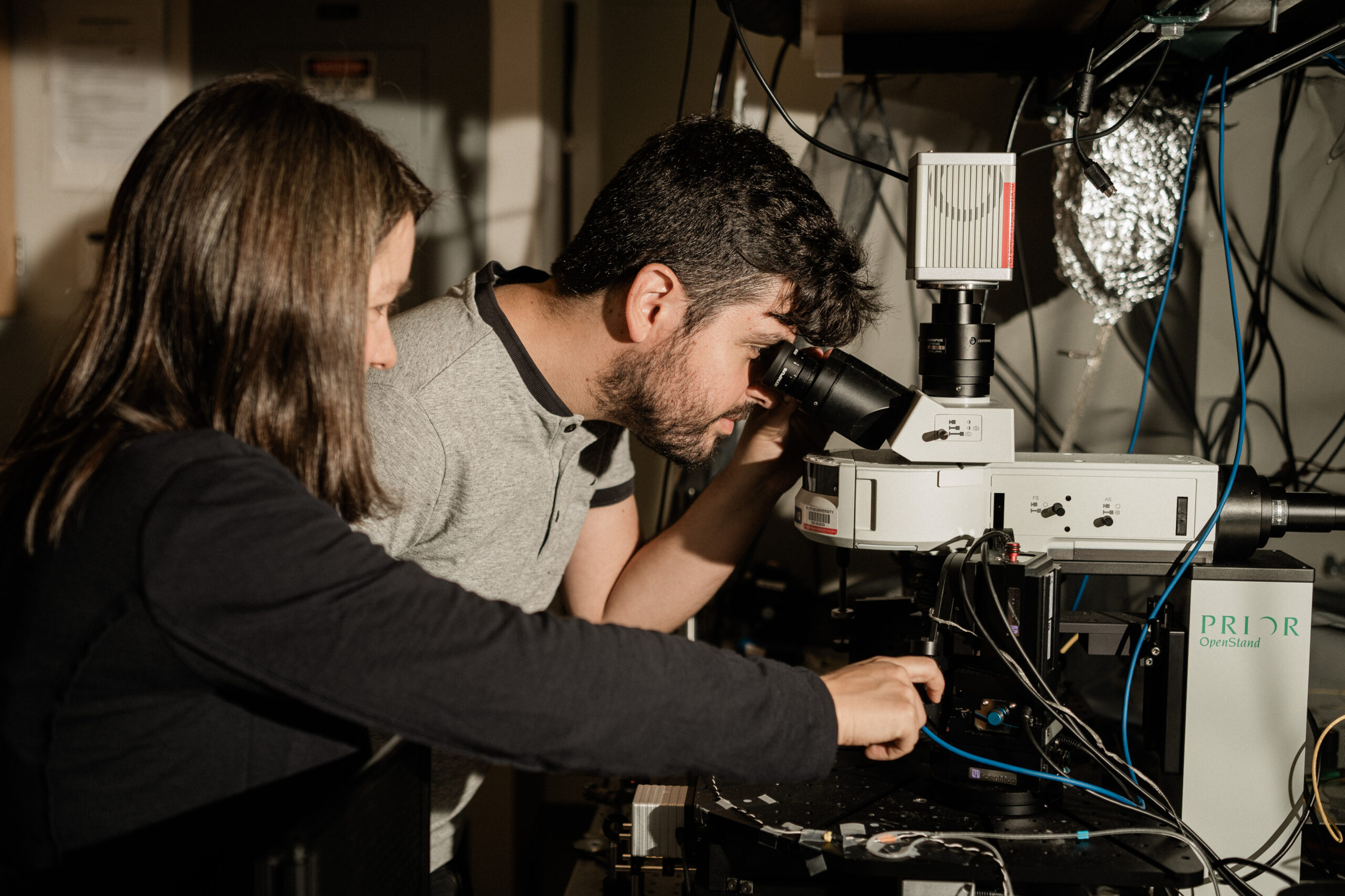
For example, the Sander group has been designing photothermal microscopes that use mid-infrared light to detect chemical signatures. As mid-infrared light is absorbed by a sample, the material is excited and heated up. Different materials feature unique characteristic responses, absorbing or losing heat more quickly or slowly depending on their chemical makeup and properties, which allows for complex structures to be analyzed and visualized. One such use for photothermal microscopy is the imaging of cells and biological structures.
But many conventional microscopes struggle to image cells and tissues inside water or other media without labeling cells with stains or fluorescent dyes. “If you have a lot of similar materials under the microscope, how do you differentiate between biological structures and the medium they’re in?” Sander says. “Water has strong background absorption at mid-infrared wavelengths, which can easily mask the signal from interesting biological features.”
By adding a shorter-wavelength laser pulse to this imaging system, Sander and her team have enhanced the spatial resolution of mid-infrared microscopy in a label-free manner.
 “This technology has allowed us to investigate chemical content in a variety of samples for biological and material science applications, as well as visualize heat transfer dynamics across a variety of interfaces,” says Panagis Samolis, a postdoctoral associate who first came to Sander’s lab as an undergraduate researcher in 2016 before staying on with the team to earn his PhD. “It’s been a very dynamic imaging technique with a lot of room to explore both optics as well as the fundamental physics of heat transfer.”
They recently used their approach to provide a closer look at axons – long, tendril-shaped parts of neurons that transmit signals to neighboring cells – in their natural watery environment. Their findings are forthcoming in Analytical Chemistry and will be highlighted on the front cover. Given that cells throughout the human body are about 70 percent water, these new capabilities could have exciting implications for biological research.
For another project, her team is working with collaborators at the Swinburne University of Technology in Australia. “We’re looking at fibroblast cells, which are these cells that form connective tissues,” Sander says.
In humans and animals, fibroblast cells make up the matrix of collagen that connects various tissues. “Using our photothermal system, we were able to image fibroblasts in their natural medium of collagen, revealing the cell and nucleus membranes on the scale of nanometers,” she says. “In our imaging technique, the fibroblasts’ cell membranes act like a thermal barrier, allowing us to better differentiate the structure of the cells and achieve heightened detail based on their heat diffusion dynamics.”
New frontiers for fiber-based laser imaging
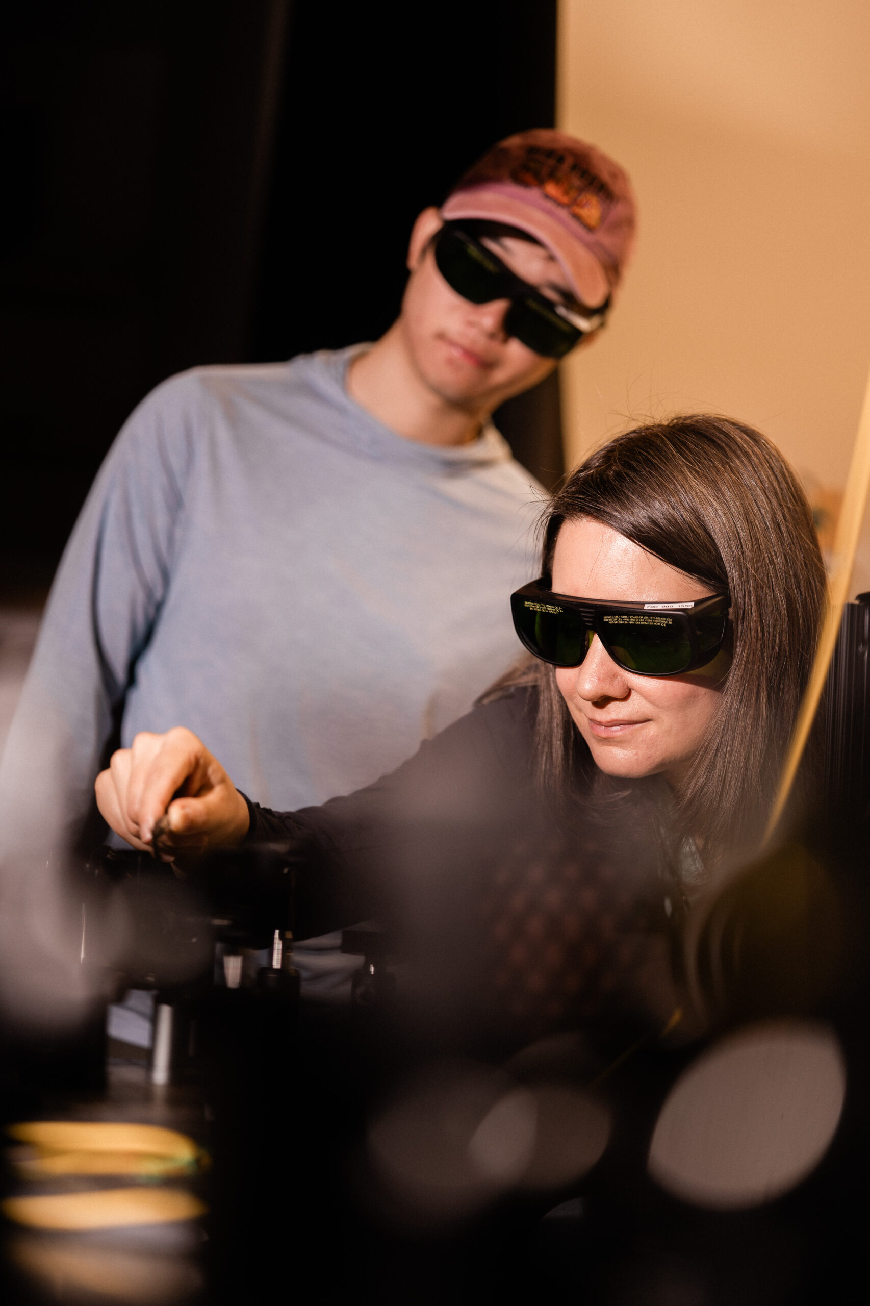
Another major focus of the lab, originally supported by an Air Force Young Investigator Award from the Air Force Biophysics program, has been developing new fiber laser techniques to probe and control the activity of neurons. Fiber lasers are extremely fine strands of silica that can transmit light from one end to the other without losing much energy and power. They are the backbone of modern-day fiber-optic telecommunications and are also an important tool used by biologists to image precise areas of biological tissues and cells.
 “We are collaborating with groups in Italy from their National Research Council in Bologna (Consiglio Nazionale delle Ricerche) and the University of Bari. They are the experts in neuronal biology, and we are the experts in imaging and probing. Together, we are working to understand how brain cells develop and communicate with each other,” Sander says.
To enable a detailed view into electrical activity between neuronal cells, and to ensure robust control while modulating brain cell activity, Sander’s team has developed fiber lasers that produce longer wavelengths (two microns) than more conventional fiber lasers.
In crayfish and other animal models, “we are using our two-micron fiber lasers to trigger neuronal activity through tiny changes in temperature caused by light,” Sander says. “Most researchers to date have focused their attention on stimulating axons, but we have instead focused on synapses,” or the junctions between neurons that send and receive electrical and chemical signals between cells. “We found that by modulating the synapse using our laser, rather than the axons, we can use less energy to induce neuronal activity.”
Ultrafast, high-energy lasers at the two-micron wavelength can also be converted to shorter wavelengths, enabling a fiber-based light source for two-photon microscopy, an imaging technique with enhanced excitation localization, improved depth, and spatial resolution than more traditional microscopy. Sander’s lab is currently working with a broader BU team to apply their amplified two-micron fiber laser system to enable two-photon voltage imaging of neuronal activity in mice.
“Ultrafast fiber lasers can be described by a single, universal math equation, but in experiments, rich, complex, and sometimes extreme or even mysterious light dynamics can be generated from these lasers,” says Shutao Xu, a fifth-year PhD student in Sander’s group. “I enjoy designing and optimizing these fiber laser systems, trying to understand each unique system’s operation in detail, and the overall process of organizing random and chaotic light into stable, coherent, ultrashort pulses. I’m very excited about seeing our lasers applied to two-photon imaging setups so that scientists can look deeper into biological tissues at a faster speed.”
Ideas, ambitions, and genuine care
In the lab and in the classroom, Sander says she is fueled by everyday moments of learning and inspiration, and by mentoring up-and-coming engineers. To date, her lab has brought more than 30 undergraduate researchers onto their team. “When you see a lab member or student struggling to make a breakthrough, and then all of a sudden they get it, and you see the light in their eyes, it’s extremely rewarding.”
She’s also recruited students from other countries, including Australia, Germany, and France, to visit and join her lab. “Creating a working environment where there is perspective from other cultures and from external institutions is very enriching for our team” she says. “I think BU has been a great place for my team to be – with all BU’s interdisciplinary centers, there are so many opportunities locally and abroad to network with colleagues from other fields. It feels unique to be in an institution that creates so many different points of contact for researchers to explore.”
Members of her team say they have developed genuine care and appreciation for their teammates in the lab and beyond work on a personal level. “In this environment, I’ve found myself able to openly share ideas and ambitions, as well as find and keep the self-motivation needed throughout my [eight-plus] years in Dr. Sander’s lab,” Samolis says. “She always leaves room for her students to find their pathways as future scientists while providing support and accommodating our individual characters, strengths, and weaknesses.”