
Issue Archives
Bostonia is published in print three times a year and updated weekly on the web.
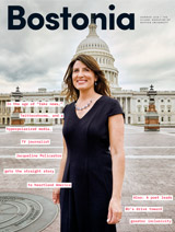
Summer 2018

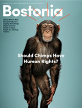
Winter-Spring 2018

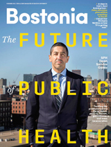
Fall 2017
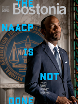
Summer 2017

Winter-Spring 2017

Fall 2016
Summer 2016

Winter-Spring 2016

Fall 2015
Summer 2015

Winter-Spring 2015

Fall 2014
Summer 2014

Winter-Spring 2014

Fall 2013

Summer 2013

Winter-Spring 2013

Campaign 2012
Summer 2012

Winter-Spring 2012

Fall 2011
Summer 2011
Winter-Spring 2011

Fall 2010
Summer 2010

Winter-Spring 2010

Fall 2009

Summer 2009
Spring 2009

Winter 2009
Fall 2008

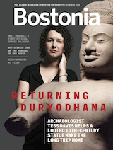
Big Shot
Billie Weiss is living out a major-league dream as manager of photography for the World Champion Boston Red Sox

Opening Doors
Erica Mosca empowers first-gen college students
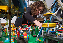
Bricks of Life
Biologist and Lego collector Cynthia Bradham finds inspiration in the tiny building blocks of life

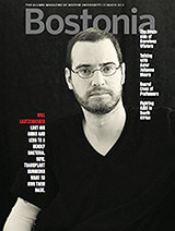
Rahul Desikan's Story of Love, Science, and Facing Down Death
This pioneering neuroscientist was attacking ALS. Then ALS attacked him.

Taking His Time
Ian Schon (ENG’12) makes analog watches in a digital world
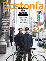
Exploring Endurance
Best-selling author David Grann (GRS’94) on one man’s obsessive quest to cross Antarctica

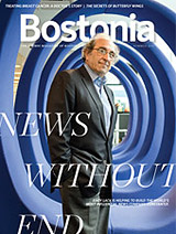
Know What’s Good for Your Health? Artificial Intelligence
Data and algorithms can spot medical concerns early and point to solutions

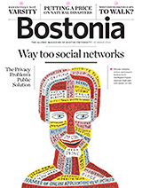
Crowdsourcing a Second Bill of Rights? ENG Professor Wants to Give It a Shot
Greg Blonder says our leaders often obstruct the public’s will

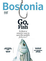
A Dead Humpback, a Team of Scientists, a Race for Answers
Did noise pollution in the ocean contribute to her death in the waters off Cape Cod? Her whale ears may hold important clues

Shari Davis (Sargent’10,’12) among 20 Obama Fellows Named Worldwide
“The Obamas represent…the reality that ceilings can and should be broken”

Natick’s World War II Museum Commemorates 75th Anniversary of D-Day
Founder Ken Rendell, who studied business at BU, has created special exhibition

Stan Sclaroff Named Dean of Arts & Sciences
After nationwide search, interim dean chosen for strong and consistent leadership
Related Stories
BU Brain Researchers among Those Invited to White House
On hand as Obama announces expansion of BRAIN Initiative
Center for Systems Neuroscience Opens in July
Will enhance interdisciplinary explorations of brain functions
Kilachand Center’s New MRI Scanner Yields Outstanding Data
CNC team invites neuroscientists across both BU campuses to use machine
Post Your Comment