The core facility is home to a variety of imaging instruments ranging from advanced laser scanning microscopes to ultrasound imaging systems. Though our central focus is supporting and using these instruments in neuroscience research, these techniques can be applied towards other aspects of biomedical imaging. On this page, you will find technical information about the various imaging systems that is available for core users.
fNIRS System I: CW-NIRS System utilizing laser diodes at 690 and 830nm (TechEn Inc. MA, USA). The TechEn system is a multichannel continuous wave optical imager consisted of 32 frequency-encoded lasers (half of them at 690 and the other half at 830 nm) and 32 avalanche photo-diode detectors.
fNIRS System II: NinjaNIRS is a BU home-built modular and fully scalable wearable NIRS device utilizing optodes with of one dual wavelength LED (730/850 nm) and one photodiode
fNIRS Head Caps: 3D printing of NinjaCaps
Fibers: Source/detector optical fibers for fNIRS System I
fNIRS Assistance
fNIRS Data Collection: Training and supervision on fNIRS data collection with both fNIRS System I and II.
Questions – Please fill out this form with any questions you may have.
Speckle/Spectral/SFDI Imaging
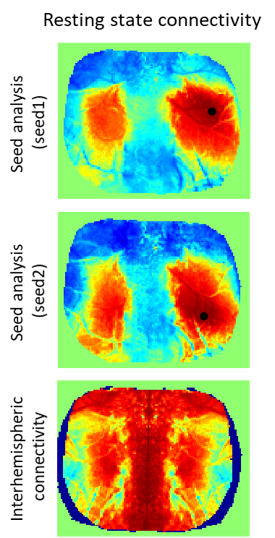
Resting state functional connectivity maps
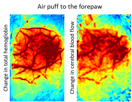
Functional activation during forepaw stimulation
Change in scattering coefficient after stroke
This is a home built wide-field microscope that can simultaneously obtain relative changes to cerebral blood flow using laser speckle contrast imaging and changes to oxy- and deoxy-hemoglobin using intrinsic optical signal imaging. Details of the combined speckle and spectral imaging system is described here. This system has also been integrated with a spatial frequency domain imaging setup based on the designs provided by Professor Roblyer’s group as described here.
Questions – Please fill out this form with any questions you may have.
Optical Coherence Tomography
We have a Thorlabs Telesto OCT system with an integrated laser speckle (LS) camera.