Participants
Click on name to see their abstract and elevator pitch video.

The purpose of this research project is to utilize micro Computed Tomography (microCT) to create 3D reconstructions of ex vivo patient tissue samples as a method of diagnosis by visualizing protein distribution in cells. Even further, the experiment tested whether or not Rolling Circle Amplification (RCA) would be an effective method of making proteins visible to CT. By conjugating a ssDNA primer to a protein specific antibody, an RCA template would be able anneal to the primer attached to the localized antibody. An X-Ray absorptive matrix would then be produced via RCA of the template to incorporate brominated/iodinated nucleotides. Such a reaction would first require antibody conjugation with a ssDNA primer. An azide transfer reaction was used to impart click reactivity to the 5’ end of the ssDNA primer using imidazole-sulfonyl azide as an azide donor. An NHS coupling reaction was used to connect a linker with a terminal DBCO moiety to the antibody. Finally, an azide-alkyne click reaction to attach the oligomer to the antibody was performed. The circularized template can then anneal to the primer which can be extended with the aid of a DNA polymerase to create an elongated nucleotide chain which forms a matrix. The brominated/iodinated nucleotides incorporated into the matrix during rolling polymerization heavily attenuate X-Rays and indicate the location of target proteins. This technique will identify targeted proteins in tissue samples and since iodinated and brominated matrices have distinctive attenuation coefficients, iodinated and brominated matrices can be simultaneously resolved, allowing for dual target imaging.

The transcription factor NF-κB plays critical, conserved roles in the regulation of genes responsible for immunity, stress response, and development in higher organisms. Nevertheless, little is known about NF-κB proteins and their related signaling molecules in more basal organisms. To learn more about the evolutionary history of these proteins, we study the sponge Amphimedon queenslandica (Aq), which has been hypothesized to be the oldest common ancestor to all animals. Aq-NF-κB contains a highly homologous region called the Rel Homology Domain (RHD), which is known in higher organisms to be responsible for DNA binding and dimerization. We aim to purify the RHD domain from A. queenslandica and determine its structure using X-ray crystallography. We hypothesize that this RHD will have a similar structure to modern day p100/p105 NF-κBs rather than Rel proteins since it is thought that the phylum Porifera to which A. queenslandica belongs branched off 630-600 million years ago, before the appearance of the first Rel proteins. Since p100/p105s have been shown to bind to DNA in homo- and heterodimers we hypothesize that the Aq-RHD will have a similar structure. As the first step towards the purification of this RHD protein, we PCR-generated an Aq-RHD truncation mutant and then subcloned this truncation into the pET28a vector, which contains a kanamycin resistant gene for selection and a HIS-tag for protein purification. For future experiments, we will express this plasmid in bacteria to extract and purify the HIS-Aq-RHD protein and prepare it for X-ray crystallography. Finding the structure of Aq-RHD can give us insight on how NF-κB proteins evolved and how they function in more basal organisms.

Morphogenesis, the patterning of tissues during animal development, is a complex and incompletely understood process. The larval sea urchin skeleton provides a tractable model of morphogenesis during animal development to investigate this process. The skeleton is formed by primary mesenchyme cells (PMCs), a migratory population of cells whose positioning determines the skeletal pattern. PMC migration is directed by patterning cues from the ectoderm. The PMCs first form a ring-and-cords arrangement to form the primary skeleton and then migrate further in order to form secondary skeletal elements. Microtubules, a component of the cytoskeleton, have been demonstrated to be involved in cell migration by establishing the polarity of the cells and maintaining the spatial and temporal coordination during migration. To assess the role of microtubules in development of the larval urchin skeleton, we treated embryos with nocodazole, an inhibitor the microtubule polymerization in high concentrations and, in lower concentrations, an inhibitor of microtubule dynamic stability. Embryos treated with nocodazole at 13 hours post-fertilization (hpf), shortly after PMC ingression into the blastocoel, developed their primary skeletons but had stunted growth and lacked certain secondary skeletal elements when compared with vehicle controls. To determine whether these defects were due to altered PMC migration, we immunostained PMCs with a PMC-specific antibody and visualized PMC position using confocal microscopy. At 18 hpf, by the end of primary PMC migration, the PMCs of the treated embryos had arranged themselves into a ring and cords but were spaced in irregular clumps. These results demonstrate that microtubule function is necessary for normal skeletal patterning and PMC migration to occur and underscores the importance of microtubules to morphogenesis in animal development.

When some materials are bombarded with an ion beam, ordered patterns form on the surfaces of these materials, and the reasons why these patterns form are largely unknown. The research topic being studied here is the stress development on silicon samples resulting from argon ion bombardment. There is not a lot of agreement among researchers around stress development on silicon samples or other surface processes that occur due to ion bombardment. Much of the available data surrounding this topic is conflicting and lacks reproducibility. So, this experiment attempts to collect more reliable data. To collect experimental data, an ultra-high vacuum chamber was used so that the experiment could be performed in a controlled environment. When bombarded, the samples bend due to the development of stress. That curvature was measured and used to calculate stress with Stoney's equation. One thing that may make this data more reliable than others is the use of a plasma bridge neutralizer, which neutralizes the ion beam hitting the sample in an attempt to minimize any curvature that may be caused by charging of the sample. We found that for angles near normal incidence, such as 0 degrees and 12 degrees, the sample experiences compressive stress which increases during bombardment, then decreases somewhat and plateaus so that the sample is experiencing moderate compressive stress for the remainder of bombardment. For angles far from normal, such as 60 degrees, the trend is the same except with tensile stress. This work could have a considerable impact in manufacturing industries. If the processes on surfaces caused by ion bombardment were well understood, ion bombardment could be a cheap and effective way to create nanopatterns on surfaces. Also, ion bombardment happens in reactors, thin film growth, and surface cleaning processes which must be controlled. There is a need in the scientific world and in these industries to better understand what happens to surfaces when they are bombarded with ions.

Heart disease is the number one cause of death in the developed world. Long non-coding RNAs (lncRNAs) are emerging as major regulators of cardiac health and disease. Gaining more insight into the specific function of lncRNAs, such as MEG3, will allow us to better understand heart disease and find innovative ways to treat it. MEG3 is a lncRNA in the DLK1-DIO3 locus. Low levels of MEG3 can be seen in aggressive cancers. Interestingly, MEG3 is abnormally down-regulated in mouse models of heart attack. MEG3 is enriched in skeletal muscle. For all muscle types, growth and regeneration requires stem cells. In stem cells, MEG3 alters the activity of the protein PRC2, an epigenetic regulator. We have shown that when MEG3 is knocked down in muscle cells in a dish, they do not mature correctly into muscle fibers. The specific function of MEG3 in muscle tissue is unknown, but we hypothesize the MEG3 lncRNA is important for muscle development and regeneration. The knockdown of MEG3 will give insight to its specific functions. The enzymes Sap1 and Aar1 were used to cut the AAV9 plasmid at specific cut sites, allowing shMEG3 to be inserted. AAV9 infects muscle tissue specifically, so this modification of the genome of AAV9 will cause this plasmid to target MEG3 in muscle cells. This plasmid can be used to study the effects of a lack of MEG3 in cardiomyocytes, and if MEG3 turns out to play an important role, this information could inform novel treatments for heart disease.

Aurofusarin is a mycelium pigment first isolated in 1937 by Julius Nicholson Ashley from the fungal plant pathogen Fusarium culmorum. Today, it is known to be a biologically active compound with potential anti-cancer capabilities. A paper published in 2018 by the Porco Group at Boston University details a 15 step process for the total synthesis of aurofusarin. However, while aurofusarin can be synthesized successfully by these methods, there is a need to reduce the number of steps. The purpose of this study was to determine a more efficient synthesis of aurofusarin. So far, three steps have been performed:
1) 2,4,5-trimethoxybenzoic acid methylation to give a benzoic methyl ester
2) Oxidation of the ester with cerium ammonium nitrate (CAN) to give a benzoquinone
3) Michael addition of a pyrone to the benzoquinone to give an adduct
The methylation reaction was found to be easily reproducible, high yielding, and very pure. On the other hand, the CAN oxidation reaction is difficult to reproduce. However, the results suggest that increasing the amount of CAN/silica gel as well as performing the reaction in a round bottom flask for better stirring (as opposed to using a vial), tends to push the reactions to go to completion faster. The Michael addition reaction had limited success, most likely due to the presence of impurities in the CAN oxidation products, which are the reactants for the Michael addition reaction. We would especially like to perform more CAN oxidation and Michael addition reactions in the future to obtain more accurate results, as well as continue in the synthesis of aurofusarin. The effective synthesis of aurofusarin can also be extended to studying the synthesis of the biologically active bis-epoxyquinol diaporine, which can serve as a barrier to the process which facilitates macrophage conversion from the M2 (repairs, decreases inflammation) to the M1 (kills, increases inflammation) phenotype.

Secure Multi-Party Computation (MPC) is a subfield within cryptography that allows parties to perform joint computations without compromising the privacy of their individual inputs. JavaScript Implementation of Federated Functionalities (JIFF) is a customizable, open-source framework for implementing complex MPC protocols and applications. My contributions to JIFF include (1) implementing a data-oblivious sorting algorithm under MPC; (2) creating a web application, BattleShips, to demonstrate JIFF’s usability; and (3) using BattleShips to develop better algorithms for checking for equality within sets of secret shared data. I optimized new algorithms that perform 99% faster than the JIFF equality function over the same data set, and then analyzed how the new algorithms would be affected by latency and bandwidth restrictions.

Tryptophan Hydroxylase (TrpH) is an important human enzyme found primarily in brain tissue that is responsible for the hydroxylation of the amino acid tryptophan, the first step in the synthesis of the neurotransmitter serotonin. Mutations in the TrpH gene that inhibit the activity of the enzyme can lead to decreased levels of serotonin, which are associated with mental disorders such as depression. Despite its physiological significance, relatively little is known about TrpH compared to the other enzymes in the aromatic amino acid hydroxylase family. New research in the Caradonna lab is focused on purifying, expressing, and characterizing the TrpH protein in a manner analogous to our studies of phenylalanine hydroxylase (PheH). Utilizing techniques such as subcloning, purification, and kinetic enzyme assays, we will gain a greater understanding of the characteristics and mechanisms of TrpH. These results will assist in further studies of TrpH and, ultimately, its implications for human health.

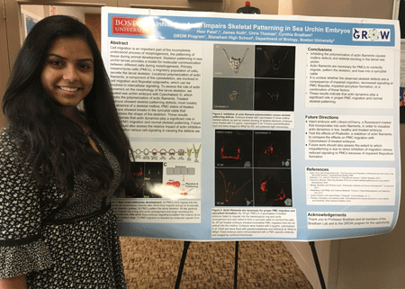
Cell migration is required for the incompletely understood process of morphogenesis, the patterning of tissue during animal development. Sea urchins are a relevant model of morphogenesis during development because their developmental patterning cues are highly conserved with those in vertebrates. Skeleton patterning in sea urchin larvae provides a model for molecular communication between different cells during morphogenesis. Primary mesenchyme cells (PMCs), a migratory population of cells, secrete the larval skeleton, and intercellular signaling from the ectoderm directss their migration. Localized polymerization of actin filaments stimulates cell migration and can change cell morphology by disassembling itself and cross-linking into bundles and networks. Furthermore, PMCs outstretch and retract filopodia, which are long, thin cellular outgrowths containing actin filaments, to the ectoderm to receive patterning information. To assess the role of actin dynamics on the morphology of the larval skeleton, we treated sea urchin embryos with Cytochalasin D, which blocks the polymerization of actin filaments. Treated embryos were imaged using transmitted light and polarized light microscopy and were also immunostained to visualize the location of their PMCs. Treatment with Cytochalasin D induces skeletal patterning defects, most notably the absence of a skeletal midline. The PMC stains of treated embryos showed losses in the syncytial cable that determines the shape of the skeleton. The PMCs inability to migrate properly and fuse together seems to have led to the lack of a syncytial cable and produced skeletal defects. These results demonstrate that actin dynamics play a significant role in proper PMC migration and normal skeletal patterning. In the future, Lifeact mCherry injected embryos can be live imaged to analyze actin dynamics in treated versus control embryos. Future work should also assess the distinction between the involvement of actin filaments in filopodia in comparison to cell migration.

There are a multitude of different methods to track cell movement. Thus far, an effective universal protocol has not been established. Especially with 3D culture systems, current software is not optimized for tracking individual cell movement. Microtissues, our platform of interest, provide a 3D culture system with a better controlled physio-chemical environment compared to animal models, and has been used to study dermal wound healing processes. Advancements in accurate cell tracking on this platform have the potential to improve our understanding of how cell movement is related to tissue remodeling and wound closure. The goal of this study is to optimize cell tracking on our platform by (a) using cells created with fluorescent markers in the nucleus and cytoplasm and (b) manipulating input parameters of cell tracking software. In this study, we successfully created fluorescence expressing cells using molecular cloning techniques, which enabled us to seed the cells into a microtissue. With fluorescent microscopy, we visualized individual cells within the tissue before, during, and after wounding. The time series of brightfield, RFP, and GFP images were analyzed using CellProfiler, an open-source image processing software, with parameter optimization steps that enhanced the software’s ability to identify and track single cells. Ongoing research explores quantitative approaches to measure both the object identification and tracking accuracy. These approaches will allow us to compare the accuracy of cell tracking between cells with only a cytoplasm fluorescent marker and cells with both a cytoplasm and nucleus marker as well as compare the effectiveness of cell tracking with different parameter inputs in CellProfiler.

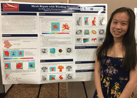
Complex meshes, used for representing 2D and 3D objects in computer graphics, often contain problematic anomalies like self-intersections and holes. These problems often prevent further processing of the mesh for rendering and other purposes. Given an input mesh description, this project repairs the mesh such that the final product is both consistent with the input shape and free of any topological anomalies. For the initial assessment of the input, the program uses winding numbers, which determine the inside or outside location of any point, to identify gaps and overlapping faces. The program then implements a segmentation algorithm using the winding number as a threshold to generate a final, repaired mesh. Since the simple threshold for segmentation often fails for low resolution meshes, the project experiments with an algorithm that determines an appropriate segmentation threshold customized for each mesh. Using the improved algorithm, this program is able to create accurate volume-based meshes from surface descriptions of objects with topological inconsistencies.

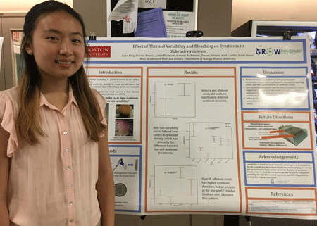
Climate change is impacting coral reefs globally, and the associated rising ocean temperatures present the biggest threat. Tropical corals, which rely on a lifelong symbiotic relationship with endocellular algae of the genus Symbiodinium for the majority of their nutrition, can lose these algae under heat stress, known as “bleaching.” The breakdown of symbiosis can lead to widespread mortality on reefs when thermal stress is sustained. Recent studies have suggested that corals exposed to more thermally variable environments may be more resistant to these bleaching events and are more likely to recover after stress. However, it is unlikely that all levels of thermal variability have the same effect on the coral-algae symbiosis. In this study, 54 coral colonies from Bocas del Toro, Panama were exposed to three different levels of daily thermal variability for 60 days followed by a stress treatment (14 days) and recovery period (14 days). To assess the response of the coral-algae partnership, densities of Symbiodinium cells present within the coral animal tissue at the end of the experiment were quantified. Preliminary results suggest that corals exposed to moderate temperature variability had the highest symbiont densities after a stress event, whereas corals that were exposed to low temperatures and other levels of thermal variability had lower symbiont densities. Our results suggest that moderate levels of thermal variation may support acclimation to stressful conditions by repeatedly exposing corals and their Symbiodinium to sublethal stress.

The primary visual cortex can utilize past experience to recognize visual sequences. The limits of this spatiotemporal learning are not well understood; for example, it is not clear how learning is affected by the amount of training. We addressed this question by performing in-vivo recordings of cortical activity in mice. Visually evoked potentials (VEPs), which are easy to record, can be used to indirectly measure the plasticity supporting spatiotemporal learning at a population level. We recorded VEPs as mice were shown a visual sequence over a course of training days. Following training, the response to the trained sequence was compared with the response to a novel sequence. The VEPs show the effect of the number of sequence training days on visual learning and prediction. The experiments show that after 2 days, potentiation occurs but it is not specific to the trained sequence. However, after 5 days, the potentiation tends to be more element specific. In addition, the data suggests that the potentiation towards the second element tends to be more stimulus specific, rather than the first element. These results indicate that spatiotemporal learning becomes more specific after a certain number of sequence training days, and that the element order is significant. The time course of stimulus specific responses constrains the mechanisms that are involved in this learning and will allow a better understanding of how neurodegenerative disorders affect the brain and its ability to learn and remember.

Surface Enhanced Raman Spectroscopy (SERS) is a field focusing on the enhancement of Raman signals, which are given off by light that scatters when a laser is fired at a target substance/molecule. Our investigations specifically focus on the distance between two components of our semi-conductive heterostructures and whether an increasing or decreasing distance has an effect on the intensity of Raman signals. Heterostructures were mainly created by stacking various materials on top of one another, all of which play an important role in the enhancement process. Silicon wafers served as our substrate, evenly coated in our target molecule, copper phthalocyanine (CuPc). Next comes the graphene, a two-dimensional sheet of carbon that served as our spacer, or the variable we’d change in order to play with the distance. Lastly, the tin (II) disulfide (SnS₂) was stacked on top, taking its place as the main signal enhancer. While most of our heterostructures took this same approach of construction, another had the positions of graphene and tin (II) disulfide switched upon stacking, which proved to be most beneficial. Being able to control the number of layers of both tin (II) disulfide and the graphene is crucial, as flakes that are either too thick or too thin will lead to weak signals. Enhancing Raman signals is a step forward in the world of substance characterization due to the valuable information obtained from this procedure, such as vibrations at the molecular level.

On a fluid-air interface, the addition of a floating particle will cause a distortion at the interface that can result in what is known as “The Cheerios Effect” where particles will cluster as a result of surface tension, just as cheerios cluster in a cereal bowl. When many particles sit on an interface, they may form a coating with varying robustness depending on the interparticle interactions. Yet, as each particle-liquid combination is unique, not much is known about their overall characteristics, such as the range of particle interactions, how the particle’s weight and shape affects these interactions, and how this contributes to the overall system strength when under strain. While past studies have explored individual fluid-particle combinations on the micro and nano scales, few have explored the concept on the macro scale with generic particles, failing to fully explain differences in the coating’s ability to withstand strain. By placing laser cut particles on a water-air interface and studying their interactions through three different methods: timing their collision at different separations, observing collision orientation in anisotropic particles, and quantifying the interface distortion through image processing, overall characteristics of fluid-particle systems were able to be observed. The study concluded that though both heavier and lighter particles are indistinguishable in visible short range effects, the heavier particles displayed more significant long-range interactions and will cluster at a faster rate. It was also found that, despite the varying distortions of an anisotropic shape, the heavier particles converged with shorter center-to-center distances, displaying the overlying effects of a monopole. As fluid-particle systems are fundamental aspects of industries ranging from cosmetics, to biomedical, to food science, understanding the fundamental principles of these systems allows for a deeper understanding for how to strengthen such systems and apply it to industry.

Quadrotors, defined as four-rotor helicopters, are becoming increasingly popular, specializing in aerial videorecording, landscape mapping, and even in a variety of humanitarian applications, such as disaster relief, first aid, search and rescue, and nature conservation. Developments in quadrotor autonomy (where a pilot or remote control is unnecessary) have also allowed for use in package delivery and human transportation (aerial taxi). However, flying a quadrotor in the real world poses many challenges: facing gusts of wind, finding flight trajectories, and autonomous decision-making, to name a few. Thus, while the demand and types of applications of quadrotors increase, so does the need for better models and controls. Therefore, in this study we examine the relation between quadrotor motor output and generated thrust. For this, I first assembled a full quadrotor, set up its software, and performed test flights on it. Then, after learning how to use related software including Python, Linux, and MATLAB, I prepared experiments, and collected and processed data off quadrotors. Finally, we determined a correlation between the quadrotor motor output and the motor rotational speed. Looking forward, this work may contribute to evolving fields such as autonomy of robots and drones, general modeling and control, and even artificial intelligence.

Urbanization around the world has led to the growth of impervious surfaces that lead to increased rates of water runoff and flooding. With the projected increase in frequency and intensity of storms due to climate change, stormwater runoff could become a greater issue in urban areas. A potential way to offset rates of runoff is to increase open, green spaces that are permeable and can naturally function as drains. However, there is not much known about what kinds of green spaces -- those with engineered soils or native soils -- are best at infiltration and preventing flooding. Because engineered soils are specifically produced to be sandy loam texture that is ideal for growing a broad range of vegetation types, I hypothesized that the texture of engineered soils would have relatively high rates of infiltration and absorbance of water compared to native soils. I collected soil samples across the Boston University campus and measured soil texture and permeability through a quantitative lab analysis using a hydrometer method. I estimated infiltration rates by using the computer program USDA ROSETTA, which converts soil texture to estimates of infiltration rates. Surprisingly, I found nearly identical rates of infiltration between native and engineered soils. Results of my research will be shared with the Sustainability Office at the Facilities Department at Boston University to build a stronger understanding of green infrastructure planning and building around our urban campus.

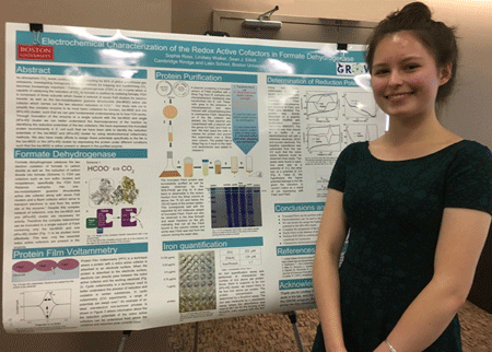
As atmosphere CO2 levels continue to rise, accounting for 82% of global greenhouse gas emissions, investigating biologically relevant methods for trapping and transforming CO2 becomes increasingly important. Formate dehydrogenase (FDH) is an enzyme which is capable of catalyzing the reduction of CO2 to formate in addition to oxidizing formate. FDH is composed of three subunits which house a network of seven FeS clusters for electron transfer as well as the bis-molybdopterin guanine dinucleotide (bis-MGD) active site cofactor which carries out the two electron reduction of CO2. Our study sets out to simplify this complex enzyme to its most basic cofactors for function, bis-MGD and one [4Fe-4S] cluster, such that we can gain fundamental understanding as to how FDH works. Through truncation of the enzyme to a single subunit with the bis-MGD and single [4Fe-4S] cluster we can better understand the thermodynamics of the reaction by identifying the reduction potentials of the two cofactors. We have expressed this truncated protein recombinantly in E. coli such that we have been able to identify the reduction potentials of the bis-MGD and [4Fe-4S] cluster by using electrochemical voltammetry methods. We also have made efforts to assign these potentials specifically to the Mo in the bis-MGD or the[4Fe-4S] cluster by expressing the protein under different conditions such that the bis-MGD is either present or absent in the purified enzyme

Quantum dots (QDs) are semiconductor nanoparticles which emit specific wavelengths of light upon excitation. These nanoparticles are of great interest for their application in solid state lighting, biosensing, and tissue depth imaging due to their tunable optical properties. InP-based QDs emerge as a non-toxic alternative to the highly characterized cadmium-based QDs. However, they are more susceptible to oxidation, have lower quantum yields, and often lose their luminescence once they are transferred into water. To render QDs suitable for future applications, excess precursors and unreacted material are removed from the reaction solution through precipitation and resuspension. This process can remove ligands from the surface of QDs and decrease their brightness. In this study, we treated three different types of InP-based core/shell QDs with fatty acids after their synthesis and purification to refine the samples for optimal fluorescence. We observe that zinc stearate significantly improves the emission counts of purified QDs for InP/ZnSe/ZnS, InP/ZnSe, and InP/ZnS quantum dots both during and after heating while the QDs lost their fluorescence after treatment with oleic acid and zinc oleate. This method can increase the emission intensity of InP-based QDs in preparation for their transfer into water, an essential step towards their biological applications.

Deoxyribonucleic acid, better known as DNA, is often referred to as one of the four molecules of life, carrying the genetic material essential for all living organisms to survive and reproduce. It has been suggested that the ability of DNA to conduct electricity has a functional role in biological processes and that it can be engineered in nanoelectronic devices as well. Numerous research groups have attempted to measure the conductance of DNA, however inconsistencies remain. In typical measurements, researchers have attached amine linkers to DNA ends to bind with gold electrodes. One reason for these discrepancies in the literature is that the question of how DNA binds to the junction remains unclear. Here we measure the conductance of two analogues of guanine, one of the building blocks of DNA, using Scanning Tunneling Microscopy-Break Junction technique (STM-BJ). Using STM-BJ, we repeatedly form and break Au-Au junctions so that, in the presence of our molecules of interest, one can bridge the Au-Au gap and we can measure the current across the Au-molecule-Au junction. We find that the guanine analogues can bind to gold and hypothesize that they are binding through the native amine and the imidazole in the guanine. This is the first use, to our knowledge, of the imidazole ligand as a binding moiety in STM-BJ single molecule conductance experiments. These measurements suggest that DNA, which contains guanine, may be able to bind to gold electrodes in the absence of a linker.