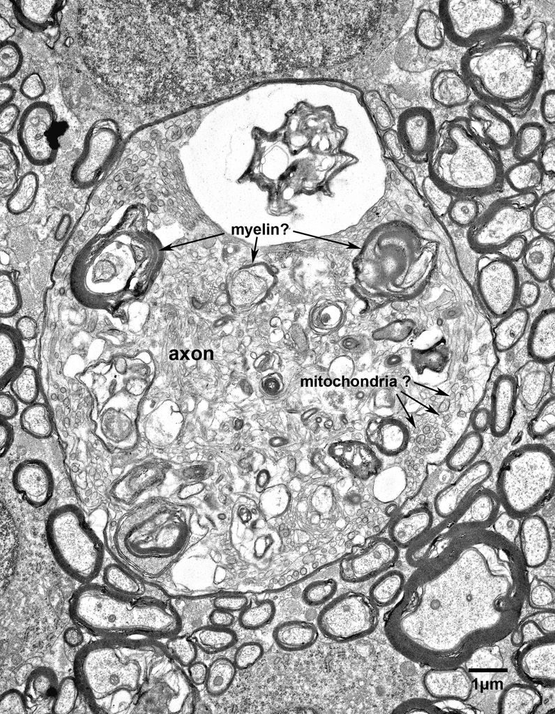
Fig. 4.4. A swollen nerve fiber in the anterior commissure of a 24 year old monkey. The cytoplasmic contents of this axon are different from the previous two examples. There are a few profiles of what appear to be modified mitochondria at the periphery of the axon, but most of the axoplasmic contents appear to be membranous, including some structures that have the appearance of pieces of myelin. This may be an early stage in the degeneration of a swollen nerve fiber.


