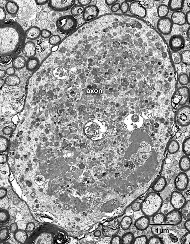
Fig. 4.2. A large swollen myelinated axon, with a diameter of about 6 microns, in the fornix of a 30 year old rhesus monkey. This type of swelling appears to result from a damming up of axoplasmic flow, causing the axon to become very swollen. This probably results in the accumulation of organelles, many of which appear to be modified mitochondria, intermixed with some lysosomes and other structures, the origins of which cannot be identified.


