NRT Trainee Poster Session
Poster Number: 1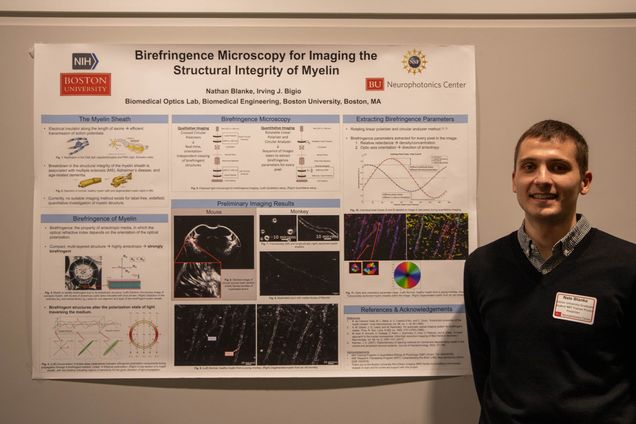
Name: Nathan Blanke
Email: nblanke@bu.edu
PI: Irving J Bigio
Department: Biomedical Engineering
Poster Title
Birefringence Microscopy for Imaging the Structural Integrity of Myelin
Abstract
Myelin is critical to neural function in vertebrates, serving as an electrical insulator along the length of axons, thus facilitating efficient propagation of action potentials. Due to the compact, layered structure of myelin, however, the study of its structure with conventional molecular labeling techniques proves to be difficult. On the other hand, by using polarizedlight microscopy to image the optical birefringence of myelin, the same properties that hinder molecular labeling can be leveraged to image details of the myelin sheath’s structural integrity. During the myelination process in the central nervous system, oligodendrocytes form projections and wrap around axons up to 60 times. (In the peripheral nervous system, the same myelination process occurs, but it is carried out by Schwann cells.) As the multi-layer myelin sheath forms, constriction displaces any cytoplasm to the periphery of the processes, resulting in a close apposition of concentric layers of lipid membrane, with the directions of the long axes of the individual lipid molecules all aligned in a radial fashion. Accordingly, the structure of the myelin sheath is highly anisotropic, and therefore strongly birefringent. Importantly, breakdown in the structural integrity of the myelin sheath has been associated with a number of neuropathies, including multiple sclerosis, Alzheimer’s disease and age-related dementia. With an initial focus on studying conditions of demyelination, we have developed a flexible birefringence-imaging microscope, with both qualitative and quantitative imaging capabilities. The system invokes two controllable, variableellipticity polarizers, one in the illumination arm, and another as the analyzer in the image-detection arm. This method of widefield microscopy uses polarized light, in a transmission geometry, to obtain dark-field, high-contrast, and label-free images of birefringent structures. The simple, yet robust, method of qualitative imaging is used for preliminary investigation of unlabeled samples in real-time, without the need for on-line image processing or scanning. For further characterization, quantitative imaging can be employed to extract the two main birefringence parameters: optical retardance and optic axis orientation, for every pixel in an image. These birefringence parameters relate directly to the molecular structural properties of the sample, to aid in identifying and quantifying morphological differences, which can inform on disease. Working with collaborators who are leading research on age-related cognitive decline, we have obtained high-resolution birefringence images of myelinated axons in brain slices from young and old primates, revealing potential age-related degeneration of the myelin sheath. Such structural information has previously only been imaged by costly and painstaking electron microscopy.
Lightning Talk Presentation:
Poster Number: 2
Name: Shuaibin Chang
Email: shuaibin@bu.edu
PI: David Boas
Department: Electrical and Computer Engineering
Poster Title
Block Face Imaging of Human Brain to Link Microscopic Immunostaining with MRI Structure
Abstract
Magnetic resonance imaging (MRI) is a non-destructive method for whole brain imaging with minimal distortions. However, current MRI techniques lack the ability to image at cellular resolution and cannot provide molecular specificity, which must be done through immuno-histology. But immuno-histology images are difficult to register back to MRI because of the inevitable distortions, tears, and warping due to tissue section and staining. Therefore, an intermediary modality must be used to help register the microscopic images to the macroscopic coordinate systems. Here, we propose a multimodal approach using block face imaging with optical coherence tomography (OCT) and two photon microscopy (TPM) to obtain pre-distorted and pre-cut cellular landmarks for registering immunostaining slices to MRI images for an entire human brain. With OCT, the intrinsic scattering and birefringence properties will allow us to use laminar structures, cytoarchitectural boundaries, and myeloarchitectural features and fibers as landmarks. With TPM, the endogenous autofluorescence such as lipofuscin provides cell body morphology for use as registration targets.
Here, we report our efforts in optimizing the image acquisition as well as characterizing the PS-OCT system and showing some preliminary data. For this task, we integrated a custom-built vibratome with a PS-OCT system, and developed software that automatically acquires the PS-OCT data. The system images a 20mm x 20mm area of agarose embedded brain tissue in 17 minutes at 10% overlap for 3mm x 3mm image tiles. In the future, the system will use a GPU to reconstruct the reflection and birefringence image on the fly. At 150um slices, our system can routinely process 20mm x 20mm x 10mm block of brain tissue in less than 24 hours.
Lightning Talk Presentation:
Poster Number: 3
Name: Timothy D. Weber
Email: tweber@bu.edu
PI: Jerome Mertz
Department: Biomedical Engineering
Poster Title
Non-Contact Phase-Gradient Corneal Microscopy with Asymmetric Transillumination
Abstract
There is significant interest in non-contact, cellular-scale corneal imaging for diagnostics and disease monitoring. Existing techniques derive image contrast from reflected light and therefore require a high degree of optical sectioning in order to reject much stronger reflections originating at other surfaces, such as the air-cornea interface. Here we present an alternative approach based on obliquely transmitted light, which produces phase-gradient contrast images of the cornea across a 1-mm field of view. Our method uses reflected light from the retina to transilluminate the cornea.
Lightning Talk Presentation:
Poster Number: 4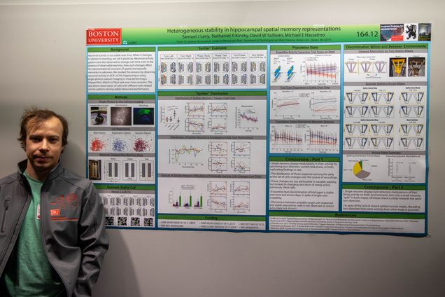
Name: Sam Levy
Email: samjl@bu.edu
PI: Michael Hasselmo
Department: Center for Systems Neuroscience
Poster Title
Representational Reorganization in Hippocampal Memory Representation in Mice
Abstract
Hippocampal cells are known to remap their firing rates and global responses to immediate changes in task demands (Wood et al., 2000, Smith et al., 2006) and to changes in the environment (Wills et al. 2005, Leutgeb et al., 2005). This results in population codes related by graded similarity for distinct experiences. Gradual changes in the representation, or “drift,” have also been observed, which don’t affect the day-to-day precision of navigation (Rubin et al., 2015). We have expanded on these findings and have shown that hippocampal representations can maintain day-to-day representational stability at the population level while being reorganized among single units. Here, we present new results describing how long-term processes of reorganization may interact with those which encode new experiences. To address these questions, we trained mice to perform multiple tasks in the same spatial environment over many days, and made recording of neural activity in CA1 using single-photon calcium imaging with head-mounted miniscopes. We then examine how long-term trends on the evolution of single neuron and population level activity are affected by performing multiple tasks in the same spatial environment, and attempt to delineate conditions under which memory reorganization is divergent at different levels of analysis. The results advance our understanding of memory maintenance and formation in the presence as an ongoing and dynamic process.
Lightning Talk Presentation:
Poster Number: 5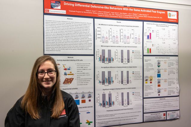
Name: Kaitlyn Dorst
Email: kedorst@bu.edu
PI: Steve Ramirez
Department: Graduate Program in Neuroscience
Poster Title
Driving Differential Defensive-like Behaviors with the Same Activated Fear Engram
Abstract
This research aims to provide a framework for the network interactions that generate differential defensive responses to aversive stimuli. Specifically, the neural circuitry that mitigates various defensive behaviors associated with post-traumatic stress disorder (PTSD) and anxiety as a result of aberrant fear memory recall are largely unknown, and these behaviors manifest as distinctive outputs depending on both the brain state and the environment of the subject. For instance, a battery of common defensive behaviors that are symptomatic of both PTSD and anxiety include, but are not limited to: active avoidance, exaggerated startle response, and freezing. Here, we alter environmental contingencies to test for the capacity of a defined set of hippocampal cells to differentially drive defensive behaviors when optogenetically activated. Our preliminary results show that artificial reactivation of the same set of cells processing fear manifests as anxiogenic responses, or freezing behavior, depending on whether these cells are stimulated during locomotion, in an open field, or in a small chamber. These results suggest that a subset of cells, upon activation, recruit different neural substrates to generate diverging behavioral outputs. Our current work utilizes immunohistochemical analyses to identify candidate regions mediating state-dependent defensive behavioral switches. Additionally, we are testing for potential interactions between artificially reactivating a fear engram during bouts of running and subsequent fear memory recall, as aerobic exercise has been shown to augment memory retention. Together, our work provides insight into the capacity of discrete sets of cells to produce varying behavioral responses.
Lightning Talk Presentation:
Poster Number: 6
Name: Sudiksha Sridhar
Email: sudiksha@bu.edu
PI; Xue Han
Department: Biomedical Engineering
Poster Title
Co-Activity Clustering Method for Quantifying Networks of Hippocampal Neurons During Learning
Abstract
Trace conditioning and extinction are two learning processes that depend on the hippocampus. We used wide field calcium imaging to record the activity of hundreds of neurons simultaneously in the hippocampus as animals were first conditioned to an aversive stimulus and then underwent extinction training to the same stimulus. We found that two distinct and separate populations of cells in CA1 of the hippocampus encode trace eye-blink learning and extinction of that conditioning. To better explore this dichotomy, we developed a novel analysis methodology based on neuron coactivity to quantify network responses across individual trials. Traditional methods based on averaging lead to loss of single trial information and are not well-suited for learning paradigms where associations can occur rapidly. Our findings suggest that co-activity clustering is adept at identifying separate clusters of neurons that contribute to conditioned stimulus encoding both before and after extinction learning. Furthermore, these features of the network also change based on whether animals exhibit the correct or incorrect behavioral response.
Lightning Talk Presentation:
Poster Number: 7
Name: Devin R. Beaulieu
Email: drbeau@bu.edu
PI: Thomas Bifano
Department: Electrical and Computer Engineering
Poster Title
Simultaneous Multiplane Imaging with Reverberation Multiphoton Microscopy
Abstract
Multiphoton microscopy (MPM) has gained enormous popularity for its unique capacity to provide high resolution images from deep within scattering tissue. We demonstrate video-rate multiplane MPM where a z-stack is acquired by near instantaneous axial scanning, while maintaining 3D micron-scale resolution. Our technique, called reverberation MPM, enables monitoring of neuronal populations over large depth ranges with no speed penalty, and can be implemented with conventional MPM as a simple add-on.
Lightning Talk Presentation:
Poster Number: 8
Name: Bahar Rahsepar
Email: baharr@bu.edu
PI: John A White
Department: Biomedical Engineering
Poster Title
Theta Phase-Specific Modulation of Engram Neurons in the Mouse Hippocampus
Abstract
Memory processing in mammalian brain is a dynamic cognitive task requiring coordination between various brain regions including hippocampus. Theta rhythm, a 4-12 Hz oscillation, is observed during memory processing. Although various behavioral studies support role of theta oscillation in coordinating activity of different brain regions, its mechanism remains unknown. A prominent computational model, Separate Phased of Encoding and Recall (SPEAR), based on in vitro electrophysiology and anatomical data, proposes different phases of theta (peak versus trough) to be involved in separating recall of stored memories from encoding of new experiences. Discovery of hippocampal engram cells, a sparse population of neurons that undergo plasticity changes during memory formation and shown to be necessary and sufficient for memory recall, has greatly advanced our understanding of brain processes underlying memory formation. Using real-time optogenetic stimulation of engram neurons, we aim to identify temporal dynamics of engram neurons, role of theta rhythm, and their attributions to formation and recall of memories. Mice were injected bilaterally with cFos-tTA and tTA-ChR2-EYFP in dentate gyrus (DG) and implanted with bilateral fiber optics in DG and a LFP electrode in the CA1 region of the hippocampus. They were exposed to fearful context A and received four electrical foot shocks. Post fear conditioning animals, were re-exposed to fearful context A as well as a novel context B. They exhibited high level freezing in context A corresponding to recall of fearful memory, while exploring naturally in context B. Later, engram neurons were optogenetically stimulated in novel context B with three different stimulation parameters: constant 20 Hz stimulation, theta peak stimulation and theta trough stimulation. Our initial results indicate peak or trough stimulation induces similar freezing levels as the natural recall in comparison to 20 Hz stimulation. Our study posits to experimentally asses proposed SPEAR model for theta by optogenetic stimulation of engram neurons in different sub-regions of hippocampal circuitry.
Lightning Talk Presentation:
Poster Number: 9
Name: Sanaya Shroff
Email: sshroff@bu.edu
PI: Xue Han
Department: Biomedical Engineering
Poster Title
In Vivo Population Voltage Imaging of Neural Activity in Awake, Behaving Mice
Abstract
A longstanding goal in neuroscience has been to image membrane voltage across a population of individual neurons in an awake, behaving mammal. Here we describe a genetically encoded fluorescent voltage indicator, SomArchon, which exhibits millisecond response times and is compatible with optogenetic control, and which increases the sensitivity, signal-to-noise ratio, and number of neurons observable several-fold over previously published fully genetically encoded reagents. Under conventional one-photon microscopy, SomArchon enables the routine population analysis of around 13 neurons at once, in multiple brain regions (cortex, hippocampus, and striatum) of head-fixed, awake, behaving mice. Using SomArchon, we detected both positive and negative responses of striatal neurons during movement, as previously reported by electrophysiology but not easily detected using modern calcium imaging techniques, highlighting the power of voltage imaging to reveal bidirectional modulation. In summary, we believe SomArchon will be easily adaptable to many contexts in neuroscience due to its fully genetically encoded nature, compatibility with simple one-photon optics and bluelight optogenetics, and ability to report report spiking activity of multiple cells simultaneously with single cell precision in awake, behaving mice.
Poster Number: 10
Name: Nicholas Vickers
Email: nvickers@bu.edu
PI: Sean Andersson
Department: Mechanical Engineering
Poster Title
Method of Synthetic Motion for Characterizing Single Particle Tracking Microscopes
Abstract
Single particle tracking (SPT) microscopes have enabled many breakthroughs in biology including understanding both molecular motor function and pathways of viral infection. A major challenge to further developing SPT microscopes is that no existing testing protocol provides a known ground truth. Simulated data have been used exhaustively in the analysis of SPT data analysis algorithms and software, however, it remains to compare different SPT microscope hardware experimentally. One method is synthetic motion, where a fluorophore fixed to a slide is moved along a realization of a stochastic motion model, such as Brownian motion, using a piezo actuated microscope stage. This motion is repeatable and known, allowing direct comparisons, and characterization of microscopes. The ability of the stage to follow the molecular motion is limited by limitations of actuator bandwidth, slew rate, and quantization. Fortunately, a feedforward-feedback control system can mitigate these allowing for a powerful tool in SPT microscope development.
Lightning Talk Presentation:
Poster Number: 11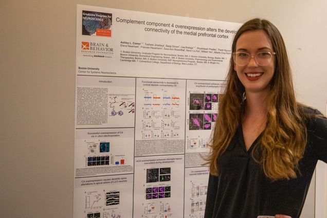
Name: Ashley Comer
Email: ashleycomer52@gmail.com
PI: Alberto Cruz-Martin
Department: Graduate Program for Neuroscience
Poster Title: Complement Component 4 Overexpression Alters the Developmental Connectivity of the Medial Prefrontal Cortex
Abstract: Recent advancements in understanding the genetic underpinnings of schizophrenia have shown increasing evidence for the role of immune molecules in the pathogenesis of this neurodevelopmental disease1,2,3,4. Specifically, the complement system, involved in innate immunity, has been shown to interfere with brain development through microglia-mediated synaptic pruning5,6. Previous work has suggested a role of complement proteins and microglia in synaptic pruning by using knock-out conditions and assaying neuronal connectivity in the visual system5,6. However, schizophrenia is characterized by the increased expression of complement proteins4. Additionally, deficits in synaptic pruning in schizophrenia are found in the prefrontal and temporal cortex of the brain7,8,9. Here, we study the role of complement component 4 (C4) overexpression in layer 2/3 pyramidal neurons in the medial prefrontal cortex of mice. Specifically, we assay neuronal connectivity by observing dendritic spine density and axonal projections. Additionally, we examine the role of microglia in altering the developmental wiring of the brain by quantifying microglia-neuron interactions and microglia engulfment in the medial prefrontal cortex. Lastly, we show that complement-induced changes to the prefrontal cortex are accompanied by deficits in social behavior in both juvenile and adult mice.
Lightning Talk Presentation:
Poster Number: 12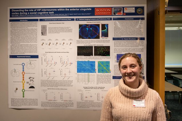
Name: Lisa Kretsge
Email: kretsge@bu.edu
PI: Alberto Cruz-Martin
Department: Graduate Program in Neuroscience
Poster Title
Dissecting the Role of VIP Interneurons within the Anterior Cingulate Cortex during a Social Cognitive Task
Abstract
As converging evidence implicates the anterior cingulate cortex (ACC) as a critical area for social cognition and emotional responses, there is an increasing need to better understand the activity of distinct cell types in this area. Vasoactive intestinal polypeptide-expressing (VIP) cells are a subtype of cortical interneurons that drive cortical disinhibition, placing them in a key position to regulate the overall activity of the ACC. Here, we investigate the role of VIP interneurons in the ACC during a task that assesses sociability and social novelty. Using miniaturized, head-mounted fluorescence microscopes (“miniscopes”) in combination with genetically encoded calcium indicators (GECIs), we are able to study the activity patterns of this sub-population within ACC in freely moving, behaving mice. We injected viral vectors containing flex-GCaMP6f, a GECI, into the ACC of VIP-IRES-Cre mice to selectively target VIP interneurons in this area. We then implanted gradient-index (GRIN) lenses in the ACC to allow for optical imaging. The miniscopes are a custom-designed variant of the open-source FinchScope. Mice were put through a battery of social and object recognition tasks to investigate the activity of VIP interneurons during social and non-social behaviors.
Lightning Talk Presentation:
Poster Number: 13
Name: Sharvari Zilpelwar
Email: sharz@bu.edu
PI: David Boas
Department: Biomedical Engineering
Poster Title
Quantifying the Impact of Scattering on the Point Spread Function Using a Modified Full Field Optical Coherence Tomography Setup
Abstract
The point spread function (PSF) is a critical parameter for all optical microscopy techniques which determines the resolution. Optical aberration is the principal source of PSF degradation, which has been traditionally corrected by specialized microscope objectives and more recently by adaptive optics. Optical scattering is another crucial source of PSF degradation in highly scattering media but the detailed effects of scattering on the PSF imaging are not experimentally explored. We developed a modified version of a full-field optical coherence tomography (FFOCT), Focused FF-OCT or 3F-OCT to image PSF degradation in the presence of optical scattering. This unique design enables measurements of unperturbed as well as the degraded PSF fields at all the depths directly. We demonstrate that the 3FOCT imaging scheme can image the PSF at the focal plane and out-of-focus planes in the weakly scattering samples. We will use this system to fully characterize the effect of scattering on the degradation of PSF. Ultimately we hope that this system will be combined with a Spatial Light Modulator (SLM) for wavefront shaping to attempt to compensate for the loss of spatial resolution due to light scattering.
Lightning Talk Presentation:
Poster Number: 14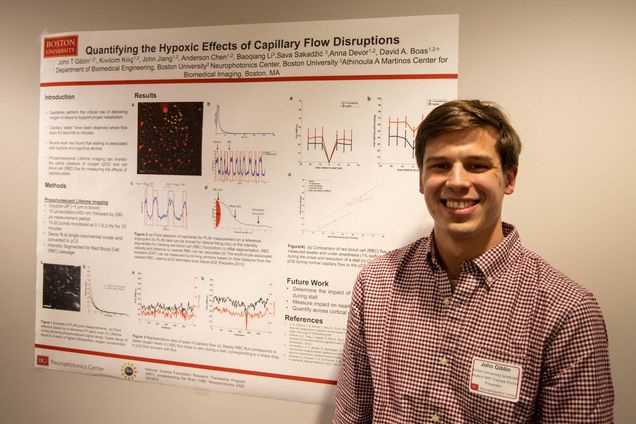
Name: John Giblin
Email: jgiblin@bu.edu
PI: Boas
Department: Biomedical Engineering
Poster Title
Quantifying the Hypoxic Effects of Capillary Flow Disruptions
Abstract
Capillaries perform the critical role of delivering oxygen to cortical tissue to support proper metabolism. Recent work has found that microcirculatory dysfunction is associated with hypoxia and cognitive decline, and potentially contributes to neurodegeneration. Specifically, capillary “stalls” have been observed where flow stops for seconds to minutes. These events are thought to contribute to changes in capillary flow distributions, which in turn inhibits the delivery of oxygen. This project seeks to understand how these transient events, along with permanent changes in microcirculatory flow contribute to cerebral dysfunction. We use Phosphorescence Lifetime Imaging to monitor the partial pressure of oxygen and red blood cell (RBC) flux in up to 20 capillaries simultaneously. During periods of stalling there was a significant observed drop in intracapillary oxygen. Our preliminary data also indicates that stalling may have a location dependent impact.
Lightning Talk Presentation:
Poster Number: 15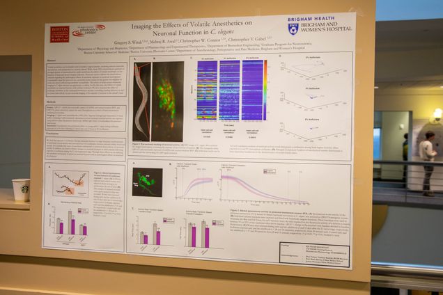
Name: Greg Wirak
Email: gswirak@bu.edu
PI: Chris Gabel
Department: Graduate Program for Neuroscience
Poster Title: Imaging the Effects of Volatile Anesthetics on Neuronal Function in C. elegans
Abstract: Volatile anesthetics are invaluable tools in modern surgical practice, rendering patients immobile, unconscious, and unresponsive to noxious stimuli. While classic EEG measurements generate an averaged picture of neuronal activity under anesthesia, the effects of volatile anesthetics on the function of neuronal circuits remains unknown. Moreover, recent evidence has raised serious concerns regarding the pathological effects of anesthetic exposure on neuronal development. Caenorhabditis elegans has proven to be a powerful model system for the genetic investigation of molecular factors influencing anesthetic susceptibility. The advent of multi-neuron functional imaging in C. elegans presents an opportunity to define, for the first time, the effects of volatile anesthetics on neuronal function with cellular resolution. We have measured the effect of isoflurane anesthetic on the command interneuron circuitry controlling crawling behavior, as well as system-wide activity via pan-neuronal imaging of the majority of neurons in the animal’s head. We find that exposure to isoflurane disrupts neuronal function via high-frequency randomization of individual neuron activity and associated loss of coordination between neurons within functional circuits. We conclude that onset of anesthesia corresponds to increased dyssynchrony of neuronal activity. In addition, we observe distinct changes in neuronal dynamics in adult animals following exposure to isoflurane during the L1 developmental stage. Through these efforts we are defining the anesthetic mechanism of action on neuronal function and its pathological effects on neuronal development.
Poster Number: 16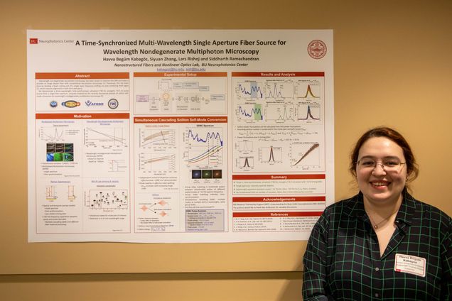
Name: Havva Begum Kabagoz
Email: kabagoz@bu.edu
PI: Siddharth Ramachandran
Department: Electrical and Computer Engineering
Poster Title
A Time-Synchronized Multi-Wavelength Single Aperture Fiber Source for Wavelength Nondegenerate Multiphoton Microscopy
Abstract
Wavelength non-degenerate two-photon microscopy has been shown to improve the SNR and make it possible to image deeper than with conventional two-photon microscopy [1]. Previously, this has been done by dividing a beam coming out of a single laser, frequency shifting one and combining them again [2], which requires alignment in both time and space. We propose to use the multi-wavelength, multi-mode output of the newly discovered soliton self-mode conversion (SSMC) [3] process for wavelength non-degenerate multiphoton microscopy. SSMC process entails one fs-pulse in a higher order mode to couple to another fs-pulse which is in a lower order mode. The frequency separation between these pulses are ~one Raman-Stokes shift and they are matched in group index, hence should be time–synchronized. Until now, a single soliton formed by the SSMC process has been shown to be time synchronized with its pump, which are generally soliton self-frequency shifted (SSFS) solitons [4]. SSMC process can also happen in a cascading fashion, meaning an SSFS pulse acts as the pump for the primary SSMC, the primary SSMC acts as the pump for the secondary SSMC, so on so forth. As a result of the cascading SSMC process, one can obtain multiple solitons, separated in wavelength –all ~one Raman-Stokes shift away from each other in frequency- and in different spatial modes. We demonstrate these cascading SSMC pulses are time-synchronized which positions them uniquely to be used as a single aperture multi-wavelength source for multiphoton microscopy.
[1] M. H. Yang, et al., “Non-degenerate 2-photon excitation in scattering medium for fluorescence microscopy,” Opt.
Express 24, 30173 (2016).
[2] E. R. Andresen, et al., “Stimulated Raman scattering microscopy by spectral focusing…,” Opt. Lett. 36, 2387 (2011).
[3] L. Rishøj et al., “Soliton self-mode conversion: revisiting Raman scattering of ultrashort pulses,” Optica 6, 304 (2019).
[4] L. Rishøj, et al., “Jitter-Free Multi-Wavelength Fiber Sources Using Intermodal Solitons,” 2019 Conference on Lasers
and Electro-Optics (CLEO), San Jose, CA, USA, 2019, pp. 1-2.
Lightning Talk Presentation:
Poster Number: 17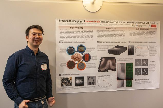
Name: Jiarui Yang
Email: jryang@bu.edu
PI: David Boas
Department: Biomedical Engineering
Poster Title
Block Face Imaging of Human Brain to Link Microscopic Immunostaining with in Vivo MRI Structure
Abstract
Magnetic resonance imaging (MRI) is a non-destructive method for whole brain imaging with minimal distortion. However, current MRI techniques lack the ability to image at cellular resolution and do not provide molecular specificity, which is typically obtained with immuno-histology. Unfortunately, immuno-histology images are difficult to register to MRI with micron-level accuracy due to the inevitable distortions, tears, and warping that arise from tissue sectioning, mounting and staining. Therefore, an intermediary modality must be used to help register the microscopic images to the macroscopic coordinate systems. Here, we propose a multi-modal approach using block face imaging with optical coherence tomography (OCT) and two photon microscopy (TPM) to obtain pre-distorted and pre-cut cellular landmarks for registering immunostaining slices to MRI images for an entire human brain. With OCT, the intrinsic scattering and birefringence properties will allow us to use laminar, cytoarchitectural boundaries, and myeloarchitectural features and fibers as landmarks. With TPM, the endogenous autofluorescence from lipofuscin provides cell body morphology for use as landmarks.
Here, we report our efforts in optimizing the tissue sample handling, image acquisition, and image-processing pipeline. For this task, we integrated a custom-built vibratome with a commercial OCT system, and developed software that automatically acquires and reconstructs OCT data on the fly. The system images a 10mmx10mm area of agarose embedded brain tissue in 20 minutes at 30% overlap for 1mm2 image tiles. With 150um slices, our system can routinely process 10x10x10mm block of brain tissue in less than 24 hours. To further improve throughput and reduce tissue damage from slicing, we will discuss the use of 2,2’-Thiodiethanol (TDE) as an index-matching agent to improve imaging depth in human brain samples. Lastly, we will report on the progress of our custom psOCT and TPM imaging microscope, which will be the next improved microscope used by this effort combined with a new vibratome to routinely image 40x40x20mm blocks of tissue.
Lightning Talk Presentation:
Poster Number: 18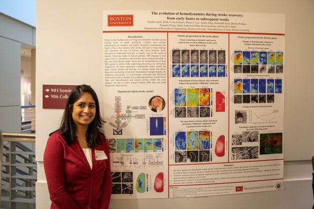
Name: Smrithi Sunil
Email: ssunil@bu.edu
PI: David Boas
Department: Biomedical Engineering
Poster Title
The Evolution of Hemodynamics During Stroke Recovery: from Early Hours to Subsequent Weeks
Abstract
Stroke is the leading cause of long-term disability and the second leading cause of death worldwide. Cellular and vascular impairments are complex and highly integrative mechanisms that begin within a few minutes after stroke and lead to tissue damage and ultimately the loss of sensory and motor function. Some spontaneous behavioral recovery is usually seen in the weeks to months following stroke in human patients. MR imaging is used extensively to characterize and monitor stroke progression both in the acute and chronic stages. However, the interpretations of these MR signals in terms of the underlying physiology are still poorly understood. We have optimized a translatable rodent model that more closely mimics the biology of a human stroke and allows long-term monitoring of recovery mechanisms through imaging. By obtaining microscopic to macroscopic structural and functional information such as changes in vascular organization, cerebral blood flow, and hemodynamic and neural activity, within the same subjects we aim to better interpret human MRI data and guide therapeutic interventions.
Lightning Talk Presentation:
Poster Number: 19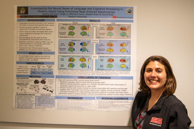
Name: Natalie Gilmore
Email: cahabjan@bu.edu
PI: Swathi Kiran
Department: Speech-Language Pathology
Poster Title
Investigating the Neural Bases of Language and Cognitive Processing in Healthy Adults Using Functional Near-Infrared Spectroscopy
Abstract
Introduction/Rationale: While functional near-infrared spectroscopy (fNIRS) is well-suited to study neuroplasticity after brain injury, it has not been applied extensively to examine language and cognitive recovery in this population to date. In order to establish fNIRS as a valuable method for studying neuroplasticity after brain injury, these processes must be thoroughly assessed in healthy younger and older adults via fNIRS as performed in this study.
Method/Approach: Twenty young (Male = 12; Age Mean [SD] = 25 [5]) and ten older, healthy individuals (Male = 5, Age Mean [SD] = 64 [6]) were recruited from the Boston area. Participants completed three tasks recruiting language and cognitive processing (i.e., semantic feature judgment [n = 20 young, 10 older]; picture naming [n = 10 young, 10 older]; arithmetic [n = 10 young, 10 older]), while undergoing fNIRS measurement. All fNIRS data was acquired using a TechEn continuous-wave NIRS device. The fNIRS probe, consisting of 56 channels, covered the following regions of interest (ROIs) bilaterally: superior frontal gyrus (SFG), middle frontal gyrus (MFG), inferior frontal gyrus (IFG), middle temporal gyrus (MTG), supramarginal gyrus (SMG) and angular gyrus (AG). fNIRS raw signal was processed and converted to concentration changes in oxygenated hemoglobin (HbO) and deoxygenated hemoglobin (HbR) via HOMER2, then, subsequently visualized via AtlasViewer using the 3-D position of reference points and optodes obtained with a 3-D digitizer. HbO concentration changes from all of the channels within one ROI were averaged across participants and run for each task. Paired t-tests were then conducted for each ROI across each time point to assess for statistically significant differences in the HbO concentration changes in the experimental condition (i.e., real pictures, or hard arithmetic) compared to the control condition (i.e., scrambled pictures, or easy arithmetic).
Results/Effects: When judging features of real versus scrambled pictures, healthy young controls showed significantly higher HbO concentration changes in bilateral MFG, MTG, AG and LIFG whereas healthy older controls revealed significantly higher HbO concentration changes in only LIFG, LMTG and RSFG. When naming real versus scrambled pictures, healthy young controls exhibited significantly higher HbO concentration changes in RIFG and RMTG in contrast with healthy older controls who showed no significant differences in HbO concentration change in any of the ROIs in this same contrast. However, healthy older controls did show significant HbO concentration changes in bilateral IFG during the real picture naming condition compared to the baseline phase. When solving hard versus easy arithmetic problems, healthy young controls demonstrated significantly higher HbO concentration changes in bilateral SFG and MFG, while healthy older controls showed significantly higher HbO concentration changes in bilateral SMG, LMFG, RSFG, RIFG and RAG.
Conclusions/Limitations: These results align with findings from previous fMRI studies using these tasks. Furthermore, they serve as a basis for understanding activation patterns in individuals with brain injury, supporting the use of fNIRS to study therapy-induced neuroplasticity in this population.
Lightning Talk Presentation:
Poster Number: 20
Name: Caroline Habjan
Email: cahabjan@bu.edu
PI: Jerry Chen
Department: Neurobiology
Poster Title
Functional and Molecular Correlates of Circuit Level Plasticity in Perirhinal Cortex
Abstract
Learning and memory are critical functions of the mammalian neocortex. On a cellular level, the modifications that accompany learning are initiated by cell-type specific, activity-dependent gene expression programs. The downstream consequences of activity-regulated gene expression result in functional changes in the way information is processed and stored in specific circuits. This project aims to identify behaviorally-relevant principles of cortical circuit plasticity in response to altered sensory experience and learning at a functional and molecular level. This will be accomplished by generating intersectional atlases of neural activity patterns and immediate early gene expression in molecularly defined cell types. Mice will be subjected to plasticity-inducing paradigms such as passive stimulation and whisker-mediated task learning. Chronic in vivo two-photon calcium imaging will be performed in perirhinal cortex to track functional changes in network activity. In a post-hoc molecular analysis pipeline, the same neurons that underwent functional imaging will be identified and will then undergo multiplexed fluorescent in situ hybridization. During which, area-specific cell-type markers and a range of activity regulated genes will be stained for, paralleling function with both stable cell-type and dynamic activity-regulated gene expression patterns on a one-to-one level. These experiments will elucidate how transcriptional profiles informs cell and circuit level activity changes that reflect plasticity, learning and memory.
Lightning Talk Presentation:
Poster Number: 21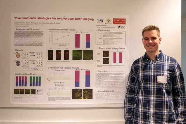
Name: Jacob Norman
Email: jfnorman@bu.edu
PI: John A. White
Department: Biomedical Engineering
Poster Title
Novel Molecular Strategies for in Vivo Dual-Color Imaging
Abstract
Calcium imaging has provided neuroscientists with the ability to monitor the activity of large networks of neurons in vivo. Novel molecular tools allow simultaneous imaging of multiple cell types, in order to better understand their real-time interactions and how they related to behavior. We compared two strategies to record simultaneously from distinguishable CaMKII-positive pyramidal cells and GAD2-positive inhibitory interneurons. In the first, transgenic strategy, GAD2- tdTomato transgenic mice were injected with jGCaMP7f expressed under the synapsin promoter. In second, double-virus strategy, AAV viral injections in GAD2-cre mice were used to drive expression of cre-dependent tdTomato and synapsinpromoted jGCaMP7f. In principle, both strategies should allow us to record from both neuronal subtypes, and distinguish interneurons based on the presence of the tdTomato signal. Our preliminary results indicate that transgenic expression of tdTomato appears to limit expression of the calcium indicator, whereas the double-virus strategy allows for greater co-expression. We have quantified the statistics in brain slices and showed that the double-virus strategy leaves around half of the GAD2+ neurons labeled with tdTomato fluorophore co-expressing jGCaMP7f. In contrast, in response to our transgenic strategy, fewer than 10% of interneurons co-expressed the calcium indicator and tdTomato. Consistent with these results, in vivo calcium recordings from double virus animals show substantially more jGCaMP7f-based activity than obtained using the transgenic strategy. These results, currently being followed up using immuno-histochemical analysis, suggest that the double-virus method is much preferred for dual-color recordings from separate, identified cell types.
Lightning Talk Presentation:
Poster Number: 22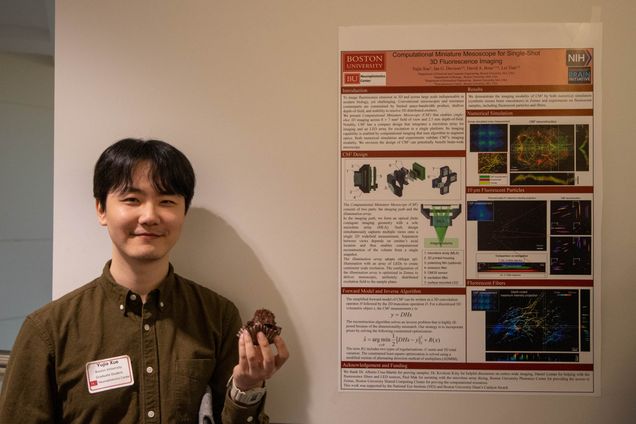
Name: Yujia Xue
Email: yujiaxue@bu.edu
PI: Lei Tian
Department: Electrical and Computer Engineering
Poster Title
Computational Miniature Mesoscope for Single-Shot 3D Fluorescence Imaging
Abstract
We report a miniature single-shot computational 3D fluorescence mesoscope. Our design is based on a finite conjugate imaging geometry with a microlens array. It achieves high resolution, depth sectioning, across a wide field-of-view in a compact design.
Lightning Talk Presentation:
Poster Number: 23
Name: Waleed Tahir
Email: waleedt@bu.edu
PI: Lei Tian
Department: Electrical and Computer Engineering
Poster Title
A Generalizable Deep-learning Approach to Anatomical Modeling of Brain Vasculature
Abstract
Analysis of brain vasculature is a fundamental problem with important applications, for which vessel segmentation is a first step. Traditional vessel segmentation methods based on parametric models have limited accuracy. More recent techniques based on machine learning have promising results but limited generalization capability. We present a deep learning based segmentation method that overcomes limitations of existing systems and demonstrates the ability to generalize to various imaging setups, samples including both in-vivo/ex-vivo data, with state-of-the-art results. We achieve so by exploiting several novel methods in deep learning, such as semi-supervised learning. We believe that our work will be another step forward towards improved large-scale neurovascular analysis.
Lightning Talk Presentation:
Poster Number: 24
Name: Johan Martinez-Fuentes
Email: jsmfuent@bu.edu
PI: Shelley J. Russek
Department: Biology
Poster Title
A Computational Model to Explore CREB-Mediated Genomic Decoding of Calcium Dynamics Resulting from Neuronal Firing Patterns
Abstract
Neuronal calcium (Ca2+) influx is important to link membrane depolarization to signaling cascade activities that regulate cellular processes including activity-dependent transcription in the nucleus. The opening of voltage-gated Ca2+ channels, induced by back-propagating action potentials (APs), is a major source of Ca2+ influx into the neuronal soma. This process can generate highly dynamic patterns of Ca2+ signaling, but the significance of this signaling pattern on gene regulatory processes remains elusive. Improved understanding may yield greater insight into mechanisms of gene dysregulation observed across many neurological disorders. An emergent idea in cell biological systems is that the genome can decode complex temporal signaling information into distinct gene expression patterns through, in part, transcription factor sensitivity and promoter activation kinetics. Here, we use in silico experiments to directly test the effect of these biochemical parameters on the transcriptional output of a prominent Ca2+ signaling-induced transcription factor, the cAMP response element binding protein (CREB), in response to a variety of empirical and simulated AP-induced Ca2+ dynamics. The computational approach combines intracellular kinetic models from the literature that preserve realistic relative rates of reaction across timescales in a simple two-compartment neuron (cytosol and nucleus). To broadly explore AP-induced transcriptional responses across long timescales, we considered experimentally measured intracellular Ca2+ traces from the literature as well as computationally inferred Ca2+ traces from simulated patterns of APs to generate pre-defined Ca2+ profiles spanning the entire simulation time is used as input. A toy set of several inducible genes, each driven by a unique promoter characterized by distinct activation rate and CREB response threshold, is implemented to generate the transcriptional output. Comparison of toy gene results with their real counterparts may suggest how certain genes may be tuned to respond to specific firing frequencies. Future studies tracking neuronal firing, dynamic CREB, and downstream transcriptional output in living cells may test model predictions. Through these in silico experiments, we address the extent by which different gene expression programs may be induced simply by adjusting the spike firing rate of a neuron. This may inform future research into gene expression changes observed in neurological disorders such as epilepsy that is characterized by abnormal bouts of hyper-synchronous neuronal excitation and calcium dynamics.
Lightning Talk Presentation:
Poster Number: 25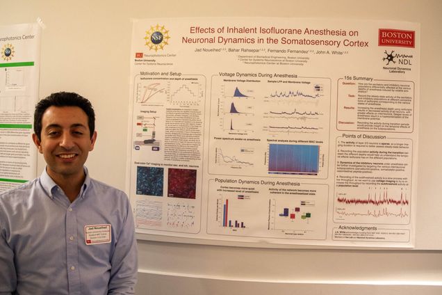
Name: Jad Noueihed
Email: jadn@bu.edu
PI: John A. White
Department: Biomedical Engineering
Poster Title
Effects of Inhalant Isoflurane Anesthesia on Neuronal Dynamics in the Somatosensory Cortex
Abstract
general anesthesia including unconsciousness, amnesia, analgesia, and akinesia. Research into how these anesthetics work has mainly progressed by characterizing either the effects on ion channels and receptors or by studying gross neural activity across brain regions. However, we still lack an understanding of how these volatile anesthetics bring about the anesthetized state and affect the network activity at the mesoscale, which is composed of tens to thousands of neurons. Bridging this knowledge gap is important as studies have shown post-operative problems linked to anesthesia exposure in infants and the elderly. To address these issues, we study the effects of isoflurane on layer 2/3 neurons of primary somatosensory cortex. Using calcium imaging we first characterize the effects on the network dynamics of the excitatory and inhibitory populations. We then use intracellular recordings to determine the effects on the sub-threshold dynamics. This study helps us understand how anesthesia is affecting the activity of the different neuronal populations, resulting in a better understanding of how the state of anesthesia is produced.
Lightning Talk Presentation: