Martin Thunemann and Ella Zeldich Receive $2.3M for Organoid Project

Assistant Professors Martin Thunemann (BME) and Ella Zeldich (Anatomy & Neurobiology) were recently awarded with a $2.3 million R01 grant from NIH (National Institute of Aging) for the next three years, with the option of a non-competitive extension to five years. The project is entitled “Understanding neuronal dysfunction in Down Syndrome using assembloids and xenotransplanted cortical organoids”.
Other co-investigators in the team include NPC associate directors Anna Devor (BME) and Christopher Gabel (Pharmacology, Physiology & Biophysics), and Joshua D. Campbell (Computational Biomedicine, Department of Medicine). This project is also in collaboration with Duygu Kuzum (ECE, UC San Diego).
This project is another example of the cross-collaborative research on organoids and assembloids, a growing research field in the STEM world––particularly in biomedical engineering. To read more on our series of organoids, see our Spring 2024 feature, or check out NPC Fellow Kate Herrema‘s research.
Below is a summary of the project narrative and abstract, provided by Drs. Thunemann and Zeldich.
Project Narrative:
People with Down Syndrome (DS) have an intellectual disability and are more likely to develop Alzheimer’s Disease (AD) earlier than the general population because of the extra copy of chromosome twenty-one, affecting neuronal activity and connectivity. In this proposal, we are using a three-dimensional patient-derived human cellular model system of regionally-patterned cortical organoids to determine how trisomy 21 affects cellular, functional, and motility-related neuronal properties and the accumulation of pathological proteins. Furthermore, our studies will include organoid xenotransplantations into mouse cortex that provides a neuronal maturation-promoting environment and allows the assessment of how spontaneous and stimulus-induced neuronal function in implanted organoids is impacted in trisomy, which will contribute to our understanding of the mechanisms underlying intellectual deficits and AD pathology in DS.
Project Abstract:
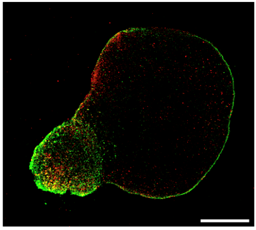
Down Syndrome (DS) is caused by triplication of human chromosome 21 (HSA21). It is a leading cause of intellectual deficits and a single genetic factor leading to the development of Alzheimer’s disease (AD). However, the mechanisms leading to cognitive dysfunction in DS are still largely unknown. While HSA21 gene triplications affect all brain cells, behavioral and cognitive deficits ultimately reflect neuronal network dysfunctions. Mechanistic understanding of altered neuronal activity is limited, as functional studies in DS individuals are not feasible and the currently available animal models for DS have considerable shortcomings. To overcome these limitations and comprehensively assess the impact of trisomy on neuronal activity and connectivity in human cells, we utilized an induced pluripotent stem cell (iPSC)-derived cellular system of cortical organoids (COs). We regionally patterned isogenic COs to generate dorsal COs (dCOs) and ventral COs (vCOs) that primarily give rise to glutamatergic neurons and inhibitory GABAergic interneurons (InN), respectively. We previously identified cellular and transcriptomic aberrations in excitatory neurons in trisomic dCOs. In our preliminary studies using calcium imaging, we detected an increased correlation of firing patterns across individual neurons in trisomic dCOs. We further expanded our experimental model and generated assembloids through the fusion of vCOs and dCOs. This powerful platform enables us to study the contribution of InN to neuronal connectivity and network activity, assess their migration as well as their role in the development of AD pathology in DS (DS-AD).
To examine the effect of trisomy on neuronal activity in a maturation-promoting environment, our team developed a multimodal recording platform where we engraft fluorescently labeled COs into the retrosplenial cortex of immunodeficient mice and monitor neuronal activity through implanted, optically transparent graphene microelectrode arrays. Xenografted COs become vascularized and establish synaptic connections with host neuronal circuits allowing us to perform longitudinal electrical and optical measurements for 6 months or longer.

Using our advanced in vivo and in vitro systems, this study will determine cellular, transcriptomic, and motility-related changes driven by trisomy and how they pertain to the alterations in neuronal activity and connectivity and the development of DS-AD. In Aim 1, we will utilize euploid and trisomic assembloids to examine the effect of trisomy on cellular composition, InN migration, and activity of individual neurons and networks using electrophysiology, calcium imaging, as well as histological and biochemical methods. Next, we will perform dCO (Aim 2) and vCO (Aim 3) xenotransplantations to address how trisomy affects (a) development of spontaneous and stimulus-triggered neuronal activity over time; (b) cellular migration and integration with the host brain; (c) transcriptomic changes within engrafted vCOs and dCOs, and (d) development of DS-AD-related features. This comprehensive panel of experiments will illuminate patho-mechanisms driving neuronal dysfunction and AD pathology in trisomy and provide a unique platform for future pharmacological, genomic, and behavioral studies.