fNIRS Comes to Boston University
The Neurophotonics Center is partnering with researchers throughout the university to incorporate the burgeoning imaging modality into their work

Dr. Helen Tager-Flusberg has made tremendous strides over the years in shedding light on language and social communication impairments in autism and other neurodevelopmental disorders. Director of the Center for Autism Research Excellence at Boston University, she has devoted herself to understanding the roots of these impairments and exploring interventions designed to help in the development of language and social communication skills.
In her work with autism Tager-Flusberg has taken advantage of behavioral and developmental approaches, study paradigms based on the observation of subjects interacting with their environment. She has also sought to probe the brain mechanisms underlying the development of language and social communication skills. Applying the tools typically used for this—neuroimaging technologies such as functional MRI—can be challenging in the populations with which she’s working. Now, though, through a collaboration with the Neurophotonics Center at Boston University, she is initiating a study with an emerging imaging modality, one that will allow her to delve more deeply into the brain and begin to uncover the many mechanisms at work.
Functional near-infrared spectroscopy—more commonly known as fNIRS—takes advantage of the properties of light to measure the amount of oxygen in the blood. Because of the tight coupling between blood oxygenation levels and activity in the brain, the technology allows researchers to monitor such activity, and in doing so begin to answer a host of questions about the brain in both health and disease. fNIRS is noninvasive and portable—imaging is done by way of a lightweight cap tethered to a larger device with fiber-optic cables, so subjects can move around and perform tasks during imaging. Thus, it can overcome some of the limitations of other neuroimaging technologies.
David Boas, director of the Neurophotonics Center and a pioneer of the technology, introduced in the mid-1990s, has been collaborating with researchers throughout the university to apply fNIRS to a broad range of studies. Because of its many unique capabilities, the approach can benefit these studies in new and exciting ways.
For example, in partnership with Boas and others in the Neurophotonics Center—including Meryem Yücel and Xinge Li —Tager-Flusberg launched a pilot fNIRS study of the mirror neuron system in autism. Mirror neurons are a type of brain cell that responds to a stimulus in the same way whether a person encounters it him or herself or simply observes others encountering it, suggesting a neurological basis for empathy. Many researchers believe that dysfunction of the mirror neuron system can negatively impact social communication skills in people with autism.
Thanks to its portability and flexibility, fNIRS opens up the study to a broader population than Tager-Flusberg could access with other imaging modalities. “I’ve done a little bit of work with much older and more verbal individuals using fMRI,” she says. “However, fNIRS offers the opportunity to look at the brain in a wider group of individuals with autism, particularly young children, who we can’t put into an fMRI scanner, and older individuals who have more challenging behavior.”
Researchers are applying the technology with other populations as well. Dr. Swathi Kiran, director of the Aphasia Research Laboratory at Boston University, studies which parts of the brain can recover function after stroke, particularly as they pertain to language. Aphasia refers to difficulty in understanding or expressing speech as a result of brain damage. In her work she has used functional MRI and other, related multimodal approaches, but measurements with these approaches can suffer from motion artifacts—a loss in data quality due to the subject moving during the measurements.
Because its design allows for motion during imaging, fNIRS is already proving an important addition to the tools used for the studies. “With functional MRI, if there’s any motion in the scanner, we’ve pretty much lost all of the data,” Kiran says. “fNIRS provides a means to circumvent some of these methodological issues, and it provides a different way of looking at the problem.”
Sharing the many benefits of the technology
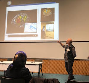
On a brisk day in January the Neurophotonics Center hosted a daylong symposium devoted to fNIRS. Held in the Kilachand Center for Integrated Life Sciences & Engineering on the BU campus, the symposium brought together fNIRS developers, users and potential users to explore best practices and the latest advances in the field. The topics covered included fNIRS as an objective measure of pain, applications in developmental neuroscience, probe design, data analysis, and more.
With six invited talks, a contributed poster session, panel discussions, a lunch and an evening reception, the symposium gave researchers interested in adopting fNIRS a wealth of opportunities to learn about the advantages the technology offers. Tager-Flusberg and Kiran both were there, as were a number of other faculty, students and postdoctoral fellows from throughout the university. All were clearly inspired by the possibilities of the technology.
“We couldn’t be more excited to introduce fNIRS to the BU community on such a broad scale,” Boas says. “The researchers with whom we’re partnering are already doing incredible work. Now, with access to this burgeoning imaging modality and the support of Neurophotonics Center staff, they can explore even further into the many problems at hand and tell us more about the brain both in healthy subjects and in a range of disorders.”