
Resting-state brain network connectivity is an independent predictor of responsiveness to language therapy in chronic post-stroke aphasia
Q&A with Ph.D. Recipient Isaac Falconer about his publication:
Falconer, I., Varkanitsa, M., & Kiran, S. (2024). Resting-state brain network connectivity is an independent predictor of responsiveness to language therapy in chronic post-stroke aphasia. Cortex. https://doi.org/10.1016/j.
Isaac Falconer, Maria Varkanitsa, Swathi Kiran, Boston University Center for Brain Recovery
Abstract
Post-stroke aphasia recovery, especially in the chronic phase, is challenging to predict. Functional integrity of the brain and brain network topology have been suggested as biomarkers of language recovery. This study sought to investigate functional connectivity in four predefined brain networks (i.e., language, default mode, dorsal attention, and salience networks), in relation to aphasia severity and response to language therapy. Thirty patients with chronic post-stroke aphasia were recruited and received a treatment targeting word finding. Structural and functional brain scans were acquired at baseline and resting state functional connectivity for each network was calculated. Additionally, graph measures quantifying network properties were calculated for each network. These included global efficiency for all networks and average strength and clustering coefficient for the language network. Linear mixed effects models showed that mean functional connectivity in the default mode, dorsal attention, and salience networks as well as graph measures of all four networks are independent predictors of response to therapy. While greater mean functional connectivity and global efficiency of the dorsal attention and salience networks predicted greater treatment response, greater mean functional connectivity and global efficiency in the default mode network predicted poorer treatment response. Results for the language network were more nuanced with more efficient network configurations (as reflected in graph measures), but not mean functional connectivity, predicting greater treatment response. These findings highlight the prognostic value of resting-state functional connectivity in chronic treatment-induced aphasia recovery.
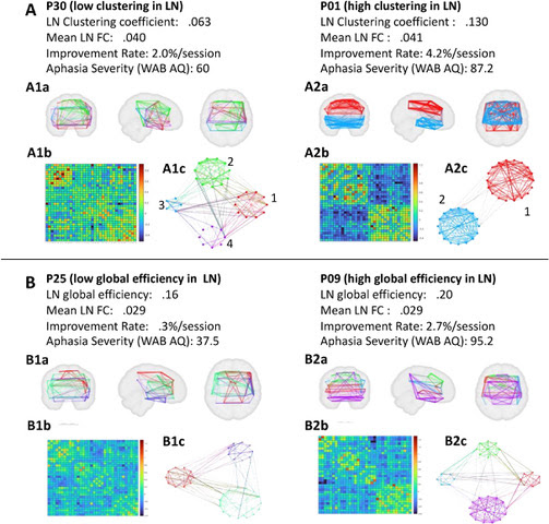
Figure 1. Individuals with post-stroke aphasia benefit more from treatment when their language network (LN) shows stronger connectivity and an organizational structure that promotes local specialization (clustering) and efficient information flow (global efficiency).
Q&A with Isaac Falconer
What is the purpose of your research?
Because individuals with aphasia follow a wide range of recovery trajectories, and treatment strategies may vary depending on the probability of benefiting from a given treatment, it is important to understand the neural and cognitive mechanisms that support treatment-induced recovery. This paper investigated resting state functional connectivity in specific brain networks as predictors of response to aphasia therapy. The results provide insights into the roles each of these networks play (or do not play) in language recovery.
How do your findings relate to the brain and recovery?
We found that greater functional connectivity in networks involved in attentional control predicted better treatment response while greater functional connectivity in a network involved in internally directed cognition (e.g., mind wandering) predicted poorer treatment response. More efficient network organization, but not overall connectivity, in the language network predicted better treatment response. Better understanding the role that specific functional networks and their associated cognitive processes play in treatment-induced language recovery will inform more effective personalized treatments for individuals with aphasia.