Mapping Recovery: Understanding Aphasia Recovery Through Neuroimaging
An Interview With 5 BU Researchers About Their Projects
In the dynamic field of cognitive neuropsychology, the integration of advanced imaging techniques with clinical outcomes offers a promising approach to understanding and treating aphasia. The Center for Brain Recovery recently completed a study that examines the functional reorganization of the language and domain-general multiple demand systems in aphasia. As part of this study, we collected structural and functional MRI data from individuals with post-stroke aphasia and healthy adults to capture detailed snapshots of the brain’s anatomy and activity patterns. We are in the process of analyzing this data in exciting new ways to investigate their relationship with behavior, providing new insights that could revolutionize therapeutic strategies. In this article, we will highlight 5 projects by graduate and undergraduate students that use this data to continue making developments in the field of recovery.
Overview of the sub-projects leveraging our data:
1. MD Network’s Role in Language Processing
Functional Reorganization of the Language and Domain-general Multiple Demand Systems in Aphasia
The study investigates potential changes in the functional architecture of the language networks in the brain and other regions not typically involved in language processing (called multiple demand regions). By expanding our understanding of language recovery beyond traditional language regions, this research could have significant implications for brain plasticity and rehabilitation, highlighting that recovery is not limited to language-specific areas. Aphasia profoundly affects quality of life, even more than cancer or Alzheimer’s disease, making this study vital for improving patient outcomes.
To learn more about this project, read our blog interview with Anne Billot from April 2023: https://www.bu.edu/cbr/2023/06/14/spotlight-research-multiple-demand-study/
2. Functional Connectivity in Diverse Language Contexts
Functional connectivity patterns in post-stroke aphasia: task level differences
Nicole Carvalho

Region of interest (ROI)-to-ROI connectivity analysis of patients vs controls, conducted in CONN.
CBR PhD. student, Nicole Carvalho, is researching functional connectivity patterns in people with aphasia while they complete different tasks, including a language localizer task, story listening, movie watching and resting-state. This study serves as a comprehensive evaluation of the overall functions and performance of the brain with aphasia.
Carvalho’s preliminary analyses show that, consistent with previous research, people with aphasia (PWA) exhibited lower connectivity than neurotypicals. “Language tasks elicited similar FC patterns in the language network, yet they elicited varying levels of connectivity in non-language networks. This variation of FC patterns may be a function of language processing that may be indicative of a PWA’s deficit or may serve to support language processing in a lesioned brain.”
3. Dynamic Functional Connectivity and Aphasia Severity
Language network dysfunction with preserved temporal variability of dynamic functional connectivity in individuals with post-stroke aphasia
Isaac Falconer
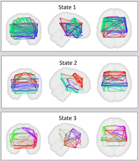
Mean dFC for time windows clustered into each of three states using k-means clustering. dFC values are indicated by line opacity. Colors indicate communities identified by the Louvain community detection algorithim. Pure colors indicate within-communities connections while internediate colors indicate between community connections (e.g., purple lines indicate connections between the red and blue communities of state)
Dr. Isaac Falconer, a recent Ph.D. graduate from the Center for Brain Recovery, explored advanced brain imaging techniques to understand post-stroke aphasia recovery. His work focused on “dynamic functional connectivity,” a method that looks at how different brain regions interact over very short times, revealing the brain’s flexibility in response to therapy. Isaac discovered that people with post-stroke aphasia (PSA) often show similar brain flexibility to healthy individuals, suggesting that their ability to improve with therapy depends more on individual differences in brain adaptability rather than the stroke’s damage itself. However, he also found that these individuals tend to have periods where their brain networks are less efficient and less well-connected, likely due to the stroke. These findings are crucial as they suggest new ways to enhance language recovery therapies by focusing on enhancing brain network efficiency.
4. Multi-modal Neuroimaging and Machine Learning to Predict Aphasia Severity
Predicting Aphasia Severity Using Multi-Modal Data
Xinyi (Selena) Hu

Feature occurrence counts for top models
Affiliated PhD. student, Xinyi (Selena) Hu, of the Center for Computing and Data Sciences, is pioneering the use of machine learning to better predict outcomes in individuals with aphasia. By integrating multiple types of brain imaging and personal demographic data—like age, time since stroke, and education level—her research aims to refine how we understand and predict the severity of aphasia. Her innovative approach combines various imaging techniques such as structural MRI, functional MRI during rest, and diffusion tensor imaging, along with sophisticated machine learning models like Support Vector Regression (SVR) and Random Forests (RF). Hu’s research shows that different modalities provide complementary information for predicting aphasia severity. The use of both SVR and RF models using combined modalities noticeably outperforms single-modality models, with resting-state functional connectivity being a key predictor. The findings of this project align with previous research, indicating that integrating multimodal neuroimaging data enhances understanding of aphasia severity and recovery (Billot et al., 2022; Chennuri et al., 2023). Hu’s work is helping further integrate machine learning and predictive modeling with the field of brain recovery.
5. Structural Integrity and Aphasia Severity:
a. Using Unsupervised Dimensionality Reduction to Identify Lesion Patterns Predictive of Post-Stroke Aphasia Severity
Emerson Kropp

Unsupervised dimensionality reduction was used on region-wise tissue-spared data from post-stroke aphasia patients to identify representative lesion patterns associated with language deficits.
Emerson Kropp, a current undergraduate student in the Center for Brain Recovery, is using neuroimaging from the dataset to analyze lesions in the brain in stroke patients, to uncover and assess their influence on aphasia. He uses a method called non-negative matrix factorization (NMF) to identify distinct patterns of brain damage and analyze how these patterns relate to the severity of aphasia. In his research, Kropp was able to identify lesion atoms that were most associated with aphasia severity, assess their pattern of damage, and compare their performance against each other. This investigation sheds light on how neuroimaging provides a deeper understanding of the causes and functions of aphasia in the brain, Kropp explains that “a better understanding of this pattern’s etiology may provide insight into the mechanisms of language dysfunction in stroke.”
b. Investigating the relationship between white matter integrity and linguistic and non-linguistic cognition in individuals with chronic aphasia
Kateri Killelea
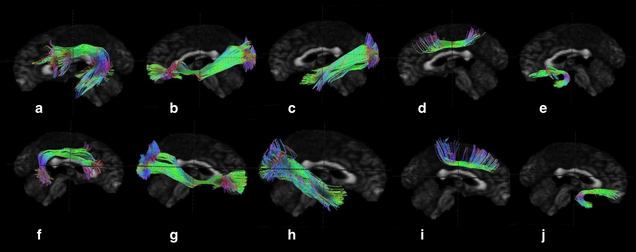
Visualization of White Matter Tracts of Interest. a- left arcuate fasciculus, b- left inferior fronto-occipital fasciculus, c- left inferior longitudinal fasciculus, d- left superior longitudinal fasciculus, e- left uncinate fasciculus, f- right arcuate fasciculus, g- right inferior fronto-occipital fasciculus, h- right inferior longitudinal fasciculus, i- right superior longitudinal fasciculus, j- right uncinate fasciculus.
CBR Master’s student, Kateri Killelea is conducting a comprehensive study on the relationship between white matter tract integrity and linguistic and non-linguistic cognition in people with aphasia. Using diffusion tensor imaging (DTI), her research builds on existing knowledge by examining key brain pathways like the arcuate fasciculus and others involved in language and cognition. Kateri’s findings confirm and extend previous research by demonstrating how the structural health of these pathways relates to specific language functions like repetition and naming, as well as to overall aphasia severity. Moreover, her work introduces new insights into the role of these tracts in non-linguistic cognitive functions such as attention, providing a more comprehensive understanding of how brain connectivity influences recovery and function in aphasia. This could lead to more targeted and effective interventions for individuals suffering from this condition.
Continuing This Research
The work being done at the Center for Brain Recovery is innovative, diverse, and developed. Students ranging from undergraduate to postgraduate levels are making significant contributions to the field with the support of the center and its mentors and leadership. In the field of recovery, neuroimaging has proved to be a key component for research, as its processing with modern computing methods and analysis will continue to bring to light important factors in how the brain functions and recovers.
To see more information on this research, subscribe to our newsletter to receive updates about our newest developments and events such as the Neuroscience in the Everyday World Conference and our CBR Seminar Series.
Our lab is continuing to grow and help students develop their research. If this work motivates you to make a contribution to support us, please visit the support our work page.