New Material for Studying Cell Mechanics May Revise Thinking on Disease
Engineering professor's research highlighted in Nature Materials
Researchers have long known that most cells living in tissues produce stabilizing networks of fibers and that the stiffness of those tissues, in turn, influences cell’s behavior. When this relationship goes awry, diseases like cancer and organ fibrosis result. But researchers have largely been studying this interaction using two-dimensional models that don’t mimic the fibrous materials in the body.
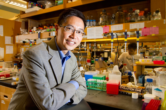
Enter Christopher Chen, a professor of biomedical engineering at Boston University’s College of Engineering, and his team, who have developed a three-dimensional platform for studying cell interactions with fibers and discovered relationships that may upend some of the thinking on how cells sense tissue stiffness. Chen and his colleagues detail their findings in a paper, “Cell-mediated fibre recruitment drives extracellular matrix mechanosensing in engineered fibrillar microenvironments,” published in October 2015 in Nature Materials.
Past research has suggested that the stiffness of the tissue that cells sit in affects their ability to spread and proliferate. Using a culture of cells grown on an elastic, flat, gel surface, previous researchers have found that as they increased the gel’s stiffness, the cells grew in size and proliferated. Recognizing that these physical conditions hardly mimicked the structure of actual tissues in vivo, Chen and his team set about creating a three-dimensional fibrous network model where the effects of tissue stiffness could be studied in a more realistic setting.
But first, they needed to develop a new material they could manipulate to adjust stiffness while mimicking the natural fiber structure of tissues. Bioengineer Brendon Baker and polymer chemist Britta Trappmann, two postdoctoral fellows in Chen’s lab, devised such a material, constructed three-dimensional matrices of fibers, embedded adult stem cells into this scaffolding, and began studying the cells’ response. Surprisingly, they found that the cells behaved very differently in these fibrous networks compared to flat gels.
For one, the pulling forces generated by cells only modestly stretched the Jell-O-like surface of the 2D model, but in contrast caused dramatic and permanent structural rearrangements in their matrix. They then adjusted the stiffness of the fibers and found that the cells—unlike those on the 2D gel—grew and proliferated more in a softer environment than in a stiffer one.
“The cell sits much like a spider within a web, applying forces to the fibers of the network,” says Baker. “These forces applied to the softer fibers allow cells to pull in or recruit more fibers, bringing more of the matrix within the cell’s reach. The cell’s ability to change its local environment could explain why we see more proliferation in this setting.”
“These findings highlight a gap in our understanding of how cells interact with their fibrous surroundings,” says Chen. “Hopefully, this new material can serve as a springboard for helping researchers elucidate the complex relationship between cells and their environment, which may bring us closer to understanding the mechanism of several diseases.”
Comments & Discussion
Boston University moderates comments to facilitate an informed, substantive, civil conversation. Abusive, profane, self-promotional, misleading, incoherent or off-topic comments will be rejected. Moderators are staffed during regular business hours (EST) and can only accept comments written in English. Statistics or facts must include a citation or a link to the citation.