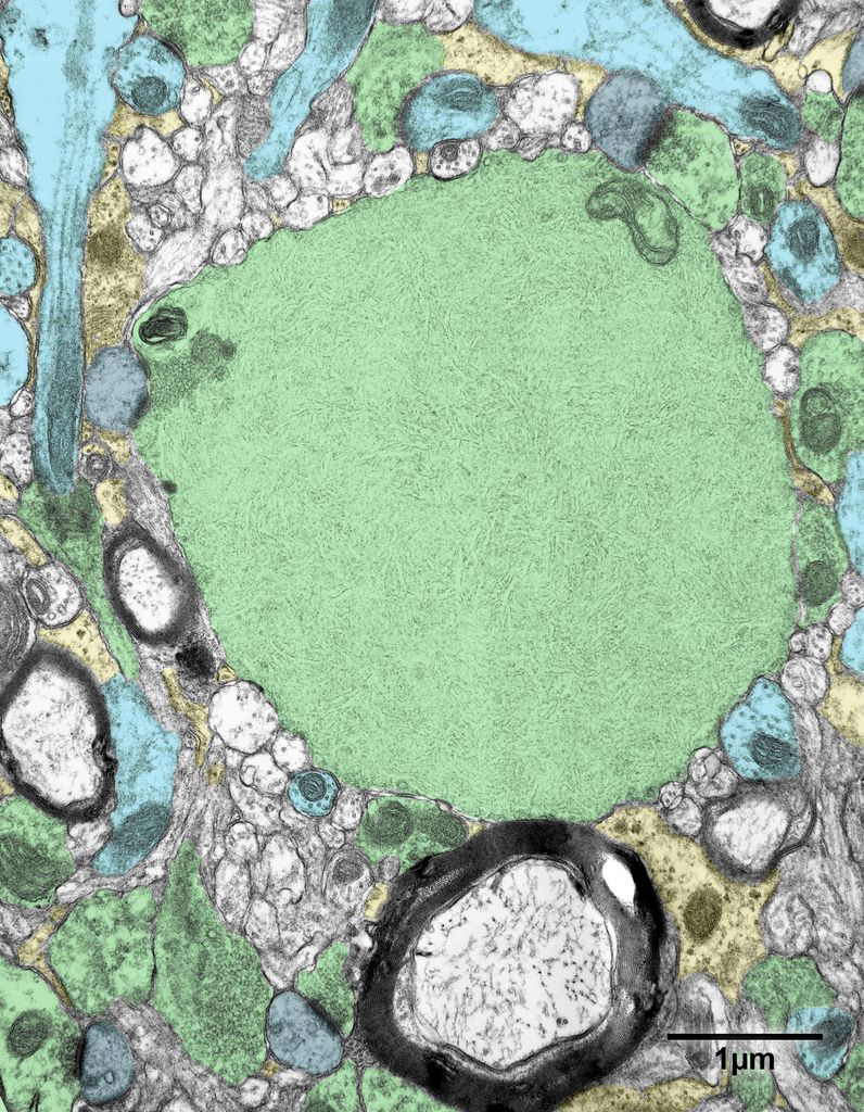
Fig. 17.3A. A colorized version of Fig. 17.3. A filamentous body in the primary visual cortex of a 35 year old monkey. The cytoplasm of this structure is filled with thin filaments, but on one side there is a cluster of synaptic vesicles where the filamentous body is forming an asymmetric synapse with a dendritic spine. Consequently this body must be an altered axon.
Filamentous body and other axon terminals- green; dendrites- blue; dendritic spines- gray; astrocytic processes- yellow.


