How Computational Imaging is Helping to Advance In-Vivo Studies of Brain Function
New BU-developed wearable device integrates miniature optics and computational algorithms to enable cortex-wide, cellular resolution imaging of the brain in freely moving animals
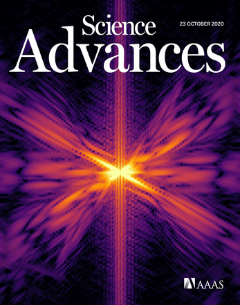
The ability to study and learn about the brain hinges on what technology is available. Current wearable brain imaging technologies are limited by a small field of view. An interdisciplinary team at Boston University led by CISE faculty affiliate Assistant Professor Lei Tian (ECE) is working to change this with the development of a novel brain monitoring system called the Computational Miniature Mesoscope (CM2). Featured on the cover of Science Advances, the CM2 will allow neuroscience researchers to advance understanding of the dynamic activity of neural circuits by allowing cellular resolution imaging of the brain across a large scale while in action.
“Neural interactions provide crucial information about the way the brain works and functions,” says Professor Tian. “Wearable devices like the CM2 aim to further the study of neural dynamics and brain activity by allowing researchers to see and study the brain through in vivo experiments with freely moving animals during naturalistic behaviors. Since functional interactions between brain areas vary with motivational state and behavioral goals, data from freely moving animals is critical.”
While there are existing technologies that image mouse brain activities, they are currently limited either by their field of view or bulky setup. Current wearable miniature fluorescence microscopes are only able to image around one square millimeter of field of view. Alternative table-top systems can provide centimeter-scale field of views, but require the mouse to be ‘head-fixed’ and secured. Since the head-fixed mouse is unable to move around, researchers are not able to gather valuable data during freely moving behaviors.
The CM2 overcomes these limitations. The device is the first of its kind, and yields a centimeter-scale field of view — 100 times larger than previous miniature microscope — while still providing single-neuron resolution. This lightweight, wearable device offers both a drastically larger field of view and built-in illumination. The larger field of view means that researchers will be able to record the neural activities across the entire brain cortical surface, rather than a small specific brain region. The high resolution ensures that researchers will be able to record neuronal activities from each neuron. In conjunction with the device’s 3D imaging capability, it promises to produce some of the most comprehensive brain images to date. The CM2 also allows for imaging to be done during movement and interactions, as the device is wearable and does not require the mouse to be secured.

The intersection of optics and machine learning for clearer, complete images
The CM2 provides expanded imaging capabilities using computational imaging that augments the optics by algorithms. The images produced by the optical component of the device on its own are composed of multiple views of the 3D object. When processed by a reconstruction algorithm, it produces a high-resolution 3D reconstruction. An array of microlenses, inspired by a compound eye, was created to allow the device to focus on the brain’s curved surface. A specially designed LED array coupled with 3D-printed light shaping apertures provide the integrated widefield illumination needed for in vivo brain recording.
“Computational imaging is imperative to understand when looking at the development of the CM2,” says Tian. “Creating such a device has proven that by utilizing coherent joint design of optics and computational algorithms, more can be done with conventional optics than has ever been done before.”
Modeling Light Behavior within Brain Tissue
Beyond the device development, this paper is the first to experimentally analyze and characterize how light scattering affects the performance of this type of miniature computational microscopy device. That analysis, which the researchers are continuing, can help them better model the light behavior within brain tissues.
“The work to develop this new device is the first-generation version and proof-of-concept,” says Tian. “With awareness of all the technical requirements the miniature mesoscope needs, we can now optimize each piece to create an even more efficient and further miniaturized version.”
Interdisciplinary team brings requisite diverse expertise
 Joining Tian in this research are Professor David A. Boas (BME, ECE) and Associate Professor Ian Davison (Biology) and CISE student affiliate Yujia Xue (PhD candidate ECE) all working under the umbrella of the College’s Neurophotonics Center
Prof. Tian, whose expertise is in computational microscopy and imaging, Davison who works to understand the neural circuits of perceptions and behaviors related to smell, and Boas who works on advancing optical, acoustic and spectroscopy neurophotonic technologies and directs the Neurophotonics Center. Boas recognized the natural teaming as Davison was developing a miniaturized version of a fluorescence microscope called a miniscope for imaging a mouse brain. The initial work to create the first proof-of-concept of the CM2 was funded by a Dean’s Catalyst Award (2018).
Award-winning Academic Recognition
The CM2 has received academic recognition by the Optical Society (OSA) and the IEEE Photonics Society. For his work on the CM2, Xue won the prestigious Emil Wolf Outstanding Student Paper Prize at the OSA Frontiers in Optics (FiO) Annual Meeting. In addition to the OSA award, Xue is a finalist for the 2020 IEEE Photonics Conference (IPC) Best Student Paper award.
Learn more about the CM2 here.
This work was supported by the National Eye Institute (NEI) (R21EY030016) and Boston University Dean’s Catalyst Award.