News
CELL-MET ERC PhD Student Jessie Song’s Nasal Swabs for Use in Covid-19 Fight Featured in New Video in The Brink
CELL-MET ERC Ph.D. student Jessie Song is developing alternative nasal swabs for use in the Covid-19 fight. Her work is featured in a new video in The Brink. Read more
CELL-MET Spring 2020 Newsletter Published
The CELL-MET ERC Spring 2020 Newsletter was recently published. Read more
CELL-MET ERC Graduate Student Jessie Song Producing Nasal Testing Swabs with 3D Printing for use During Covid-19 Pandemic
CELL-MET ERC graduate student Jessie Song, who works with CELL-MET Professors Alice White and Mark Grinstaff, has produced nasal swabs with 3D printing to help alleviate supply chain problems with testing during the Covid-19 pandemic. Read more
CELL-MET ERC Students Achieve Level of Finalists in Termis Competition
Students from the CELL-MET ERC competed in the Tissue Engineering and Regenerative Medicine International Society (TERMIS) business plan competition and achieved the level of finalist. They will move on to the finals at Princeton University on April 4, 2020. Read more
CELL-MET Fall 2019 Newsletter
Our first CELL-MET ERC Newsletter is live here: CELL-MET Fall 2019 Newsletter
Directors
A swifter way towards 3D-printed organs (Wyss Institute press release featuring Jennifer Lewis)
This video shows the SWIFT bioprinting process, including forming dense organ building blocks of living cells, printing and evacuating of sacrificial gelatin ink, and creating cardiac tissue that successfully beats like a living heart over a seven-day period.
A swifter way towards 3D-printed organs from September 6, 2019.
Sacrificial ink-writing technique allows 3D printing of large, vascularized human organ building blocks
By Lindsay Brownell
(CAMBRIDGE, Mass.) — 20 people die every day waiting for an organ transplant in the United States, and while more than 30,000 transplants are now performed annually, there are over 113,000 patients currently on organ waitlists. Artificially grown human organs are seen by many as the “holy grail” for resolving this organ shortage, and advances in 3D printing have led to a boom in using that technique to build living tissue constructs in the shape of human organs. However, all 3D-printed human tissues to date lack the cellular density and organ-level functions required for them to be used in organ repair and replacement.

Now, a new technique called SWIFT (sacrificial writing into functional tissue) created by researchers from Harvard’s Wyss Institute for Biologically Inspired Engineering and John A. Paulson School of Engineering and Applied Sciences (SEAS), overcomes that major hurdle by 3D printing vascular channels into living matrices composed of stem-cell-derived organ building blocks (OBBs), yielding viable, organ-specific tissues with high cell density and function. The research is reported in Science Advances.
“This is an entirely new paradigm for tissue fabrication,” said co-first author Mark Skylar-Scott, Ph.D., a Research Associate at the Wyss Institute. “Rather than trying to 3D-print an entire organ’s worth of cells, SWIFT focuses on only printing the vessels necessary to support a living tissue construct that contains large quantities of OBBs, which may ultimately be used therapeutically to repair and replace human organs with lab-grown versions containing patients’ own cells.”
SWIFT involves a two-step process that begins with forming hundreds of thousands of stem-cell-derived aggregates into a dense, living matrix of OBBs that contains about 200 million cells per milliliter. Next, a vascular network through which oxygen and other nutrients can be delivered to the cells is embedded within the matrix by writing and removing a sacrificial ink. “Forming a dense matrix from these OBBs kills two birds with one stone: not only does it achieve a high cellular density akin to that of human organs, but the matrix’s viscosity also enables printing of a pervasive network of perfusable channels within it to mimic the blood vessels that support human organs,” said co-first author Sébastien Uzel, Ph.D., a Research Associate at the Wyss Institute and SEAS.
The cellular aggregates used in the SWIFT method are derived from adult induced pluripotent stem cells, which are mixed with a tailored extracellular matrix (ECM) solution to make a living matrix that is compacted via centrifugation. At cold temperatures (0-4 °C), the dense matrix has the consistency of mayonnaise – soft enough to manipulate without damaging the cells, but thick enough to hold its shape – making it the perfect medium for sacrificial 3D printing. In this technique, a thin nozzle moves through this matrix depositing a strand of gelatin “ink” that pushes cells out of the way without damaging them.

When the cold matrix is heated to 37 °C, it stiffens to become more solid (like an omelet being cooked) while the gelatin ink melts and can be washed out, leaving behind a network of channels embedded within the tissue construct that can be perfused with oxygenated media to nourish the cells. The researchers were able to vary the diameter of the channels from 400 micrometers to 1 millimeter, and seamlessly connected them to form branching vascular networks within the tissues.
Organ-specific tissues that were printed with embedded vascular channels using SWIFT and perfused in this manner remained viable, while tissues grown without these channels experienced cell death in their cores within 12 hours. To see whether the tissues displayed organ-specific functions, the team printed, evacuated, and perfused a branching channel architecture into a matrix consisting of heart-derived cells and flowed media through the channels for over a week. During that time, the cardiac OBBs fused together to form a more solid cardiac tissue whose contractions became more synchronous and over 20 times stronger, mimicking key features of a human heart.
“Our SWIFT biomanufacturing method is highly effective at creating organ-specific tissues at scale from OBBs ranging from aggregates of primary cells to stem-cell-derived organoids,” said corresponding author Jennifer Lewis, Sc.D., who is a Core Faculty Member at the Wyss Institute as well as the Hansjörg Wyss Professor of Biologically Inspired Engineering at SEAS. “By integrating recent advances from stem-cell researchers with the bioprinting methods developed by my lab, we believe SWIFT will greatly advance the field of organ engineering around the world.”

Collaborations are underway with Wyss Institute faculty members Chris Chen, M.D., Ph.D. at Boston University and Sangeeta Bhatia, M.D., Ph.D., at MIT to implant these tissues into animal models and explore their host integration, as part of the 3D Organ Engineering Initiative co-led by Lewis and Chen, both of whom are faculty members of the CELLMET center at Boston University.
“The ability to support living human tissues with vascular channels is a huge step toward the goal of creating functional human organs outside of the body,” said Wyss Institute Founding Director Donald Ingber, M.D., Ph.D., who is also the Judah Folkman Professor of Vascular Biology at HMS, the Vascular Biology Program at Boston Children’s Hospital, and Professor of Bioengineering at SEAS. “We continue to be impressed by the achievements in Jennifer’s lab including this research, which ultimately has the potential to dramatically improve both organ engineering and the lifespans of patients whose own organs are failing,”
Additional authors of the paper include John Ahrens, a current graduate student at the Wyss Institute at Harvard University and Harvard SEAS, as well as former Wyss Institute and Harvard SEAS members Lucy Nam, Ryan Truby, Ph.D., and Sarita Damaraju. This research was supported by the Office of Naval Research Vannevar Bush Faculty Fellowship, the National Institutes of Health, GETTYLAB, the NSF-CELLMET Center at Boston University, and the Wyss Institute for Biologically Inspired Engineering at Harvard University.
Center Director David Bishop highlighted in BU Today
The following is an except from the article "Meet Seven BU Researchers Who are Transforming the Human Experience" published by BU Today on Friday, September 20, 2019. Article by Kat J. McAlpine.
David Bishop, who’s developing living “Band-Aids” to repair hearts damaged by heart attack, is keenly aware of the impact that his team’s work stands to have.
“Heart disease kills one in four Americans, and it’s one of the leading causes of death around the world,” says Bishop, director of BU’s CELL-MET, an engineering research center in cellular metamaterials funded by the National Science Foundation. “A lot of us are going to die from heart disease, because right now, heart attacks can’t be cured,” he says.
After a heart attack, the muscle cells that help the heart contract die off and are replaced by stiff, immovable scar tissue. “You’re permanently disabled,” Bishop says. “When heart muscle dies, your heart can’t contract as strongly and can’t eject as much blood as you did before. Cardiologists today never use the word cure; they only talk about managing your condition.”
His vision? “If someone has a heart attack, we create new cardiac tissues derived from that person’s own cells,” he says. “We create an implantable tissue patch that covers the damaged part of the heart, to restore the heart’s capacity to pump blood. The tissue patch transmits electrical signals and contracts in a twisting and squeezing motion, just like healthy heart does on its own. No foreign organ tissues involved, no immunosuppression issues.”
The CELL-MET team is 2 years into a 10-year program designed to bring the vision of a living, beating heart Band-Aid to life. Bishop and Alice White, an ENG professor and chair of mechanical engineering, bring their nanotechnology expertise to the table. CELL-MET deputy director Christopher Chen, an ENG professor of biomedical engineering and a William Fairfield Warren Distinguished Professor, is an expert in microchip environments that mimic the inside of a living heart. These so-called “hearts on a chip” set the essential stage that lab-grown cardiac tissues need to develop and function normally. And renowned imaging scientist Thomas Bifano, an ENG professor of mechanical engineering and director of BU’s Photonics Center, is enabling the team to see and measure every aspect of heart tissue growth, down to inconceivably small cellular parts.
“Together, leveraging our strengths, we have a shot,” Bishop says.
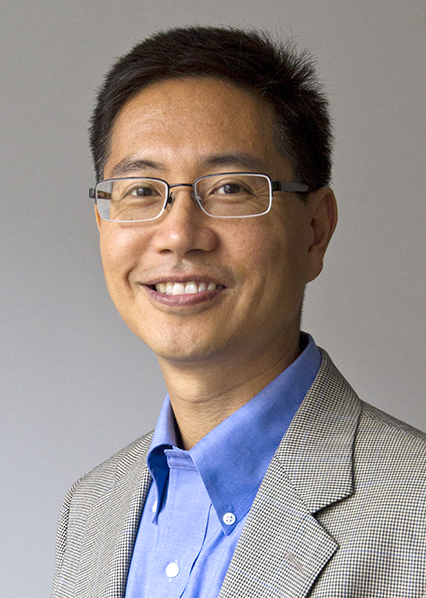
Christopher Chen honored as 2019 William Fairfield Warren Distinguished Professor
Last week, Boston University President Robert A. Brown announced "...the appointment of four of our colleagues as William Fairfield Warren Distinguished Professors. The Warren Professorship is the highest recognition that the University bestows on a faculty member. Christopher Chen, Michael Hasselmo, Xuefei Jin, and Ann McKee have been chosen to join this group, bringing the number of active Warren Professors to 13."
The following is an excerpt from a news story on BU Today by Amy Laskowski. The full article can be read here.
"The William Fairfield Warren Professorships, named in honor of BU’s first president, were established in 2008 to recognize BU’s most distinguished faculty. The award is the highest distinction bestowed upon senior faculty members who remain actively involved in research, scholarship, teaching, and the University’s civic life. It comes with an annual scholarly allowance of $20,000 and funding for a month of summer salary. Each recipient is bestowed an emeritus title upon retirement...
Christopher Chen is one of the world’s leading experts on regenerative medicine. He studies tissue engineering and mechanobiology, which combines engineering and biology to study how physical forces and changes in cell or tissue mechanics affect development, physiology, and disease. He is director of BU’s Tissue Microfabrication Laboratory, founding director of the Biological Design Center, and deputy director of the National Science Foundation Engineering Research Center for Cellular Metamaterials, both housed at Boston University. Chen is also a member of the Harvard Wyss Institute for Biologically Inspired Engineering.
“It means a great deal to be sharing this esteemed professorship with other luminaries here at BU, and especially being the first ENG faculty to be selected,” says Chen, who also recently won the 2019 Robert A. Pritzker Distinguished Lecture Award and is a fellow of the American Institute for Medical and Biological Engineering. “It is especially meaningful to know that my research and educational activities are being amplified by this honor, and I hope to continue to add to its luster.”
Chen says the news of the professorship caught him off guard. Brown had invited him to a meeting at his office “to discuss nominations for awards, so I presumed he was asking me for input on one of his projects,” he says. “I thought something was up when he and Jean Morrison sat me down and they were beaming. It was meaningful to receive the honor personally from them.” "
ENG Professor Christopher Chen at a laboratory, Cummington Mall.
Photo by Chitose Suzuki for Boston University Photography
CELL-MET member Bioventus advances bone formation for improved healing
Bioventus, a CELL-MET member, has developed a next-generation chimeric bone morphogenetic protein BMP/carrier engineered for more potent/efficaious bone formation, that acts as an adjunct to surgical repair of severe open fractures requiring augmented healing. This research article shows that a BMP/activin A chimera is superior to native BMPs and induces bone repair in nonhuman primates.
For more information on this study, please visit www.ncbi.nlm.nih.gov/pubmed/31019025
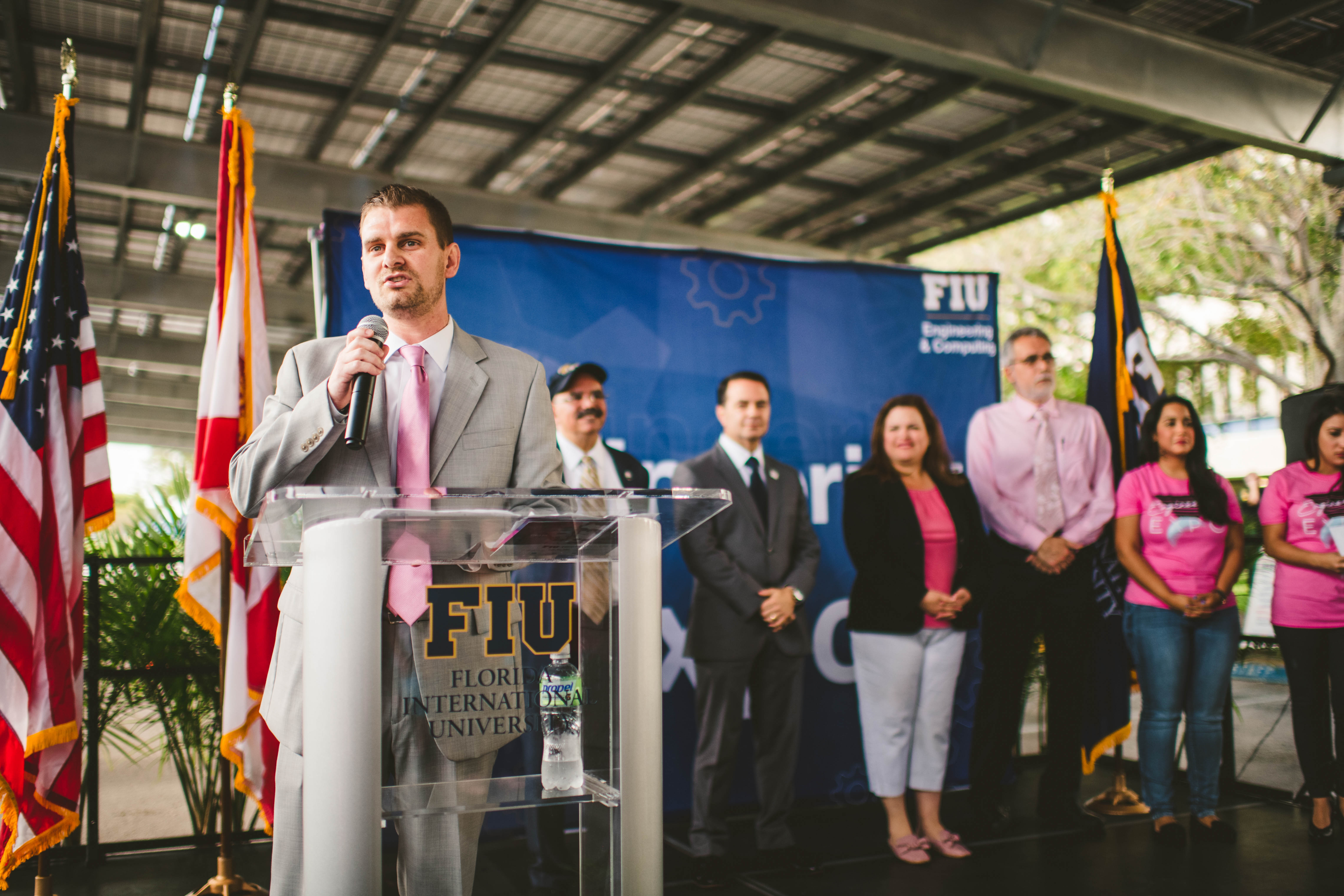
Engineering Expo at FIU inspires youth interest in STEM studies
Click here for a video highlight of the 2019 Engineering Expo at FIU
On February 22nd, 2019, Florida International University hosted hundreds of local grade schoolers at the 2019 Engineering Expo. The event began with a welcome address from John Volakis, Dean of Engineering & computing at FIU, as introduced by CELL-MET Culture of Inclusion team member Andrew Green. Throughout the day, students went from station to station, learning about different engineering projects at FIU. Engineering Workforce Development and Education students Justin Fitch, Cami Madera, and Javier Garcia lead a station focused on CELL-MET’s nanotechnology and the fascinating goals of the ERC with regards to tissue engineering. Included in the rotation was a tour of the Nanobioengineering / Bioelectronics Laboratory. The purpose of the expo was to spark interest in STEM research among the local youth, hopefully inspiring their future studies in these areas of education.