Return to Woundbiotech Home Page
Return to Growth Factors
I. Introduction
Over the past several decades, the discovery of growth factors has led to much hope and speculation about the use of these potent peptides in the treatment of difficult to heal wounds, particularly chronic wounds. In vitro experiments showed that growth factors were very effective in regulating cell proliferation, chemotaxis, and extracellular matrix formation. Animal experiments confirmed the notion that growth factors could accelerate wound repair, although most such experiments dealt with wounds created by acute injury. However, it was not until later, when further advances in recombinant technology made it possible to obtain large amounts of purified growth factors, that these agents could be tested in human clinical trials. Over the last 10 to 15 years, a large number of trials have been performed to evaluate the safety and effectiveness of growth factors in the healing of chronic wounds due to pressure (decubitus ulcers), diabetic neuropathy, and venous insufficiency (1). Platelet-derived growth factor (PDGF) is now approved for topical treatment of diabetic neuropathic ulcers (2). In this brief discussion, we will review growth factors, their mode of action, and the experience from clinical trials, with particular emphasis on the use of PDGF in diabetic foot ulcers. We will end our discussion by providing a perspective on the future of growth factors in chronic wounds, including diabetic foot ulcers.
2. General aspects of growth factors
Although more easily conceptualized by the division into three distinct phases (inflammation, fibroplasia, and maturation), the process of wound repair is characterized by a series of complex cellular and molecular events with a great degree of overlap and interdependence (3). Growth factors play fundamental roles in this process, by stimulating chemotaxis and cellular proliferation, by providing signaling among cells of the same and different type, by controlling extracellular matrix formation and angiogenesis, by regulating the process of contraction, and by re-establishing tissue integrity. As soon as blood vessels are disrupted, platelets enter the wound in great numbers and release several growth factors, including platelet-derived growth factor (PDGF) and transforming growth factor-ß1 (TGF-ß1). These and other growth factors are chemotactic for a number of cell types critical to the repair process, such as macrophages, fibroblasts, and endothelial cells. Later, during the proliferative phase of wound repair, several growth factors, including vascular endothelial growth factor (VEGF), fibroblast growth factors (FGFs) and PDGF and TGF-ß isoforms, provide a potent stimulus for angiogenesis and for fibroblasts to synthesize key extracellular components (i.e., collagens, proteoglycans, fibronectin, elastin). During the later stages of wound repair, growth factors are important in tissue remodeling, aided by the action of matrix-degrading metalloproteinases (MMPs). It is likely, however, that the action of growth factors does not end with wound closure and tissue remodeling, but that they are key players in the maintenance of tissue integrity and in cell to cell communication.
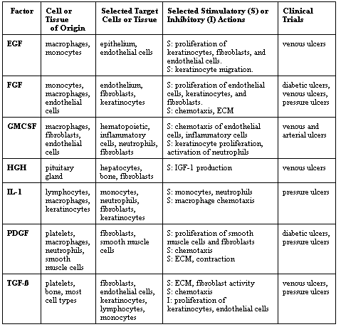
Growth factors are multifunctional peptides that are extremely potent in vitro, often in the picogram range (4). Table 1 shows a list of growth factors, some of them grouped into families, which have been tested for the treatment of chronic wounds. This list is not meant to be inclusive, and many more clinical studies have been performed than suggested by the number of published results. The nomenclature used to define growth factors tends to be confusing to the clinician for a number of reasons. First of all, the names of growth factors have more to do with the circumstances in which they were identified than with their specific effects on cells. Thus, fibroblast growth factors (FGFs) are very potent angiogenic factors, and transforming growth factor-ß's (TGF-ß's) are not transforming to cells and actually appear to be important in preventing cancer. Also, the term growth factor is generally used in a broad sense, to indicate substances which increase cell proliferation, mitogenic activity, and extracellular matrix formation. The actual category in which such substances are placed often depends on the context in which they were discovered. For example, chances are that the same substance identified by a biochemist, an immunologist, and a hematologist would be called a growth factor, an interleukin, and a colony stimulating factor, respectively.
Growth factors work by binding to specific cell surface receptors and can target cells in a number of recognized ways or modes. Release of these substances into the blood stream allows them to get to distant targets (endocrine mode). From the cell of origin, growth factors can diffuse over short distances to affect other cells (juxtacrine mode), and to influence neighboring cells (paracrine mode). Growth factors can also act on the cell in which they are produced (autocrine mode). These different modes are all likely to be operative during tissue repair (1,4).
After binding to receptors, growth factors can have a profound influence on cell proliferation, chemotactic activity, and extracellular matrix synthesis. Interestingly, not all of these actions are stimulatory and not for all cell types. For example, transforming growth factor-ß's (TGF-ß's) are potent inhibitors of keratinocyte and endothelial cell proliferation. However, these same agents are a potent stimulus for the deposition of collagen and other extracellular matrix proteins. Harvesting the inhibitory activities of growth factors has great therapeutic potential, i.e., using antibodies to TGF-ß's to decrease scarring.
3. Mode of action of growth factors
Much of the progress made in the last ten years in the basic science aspects of growth factors has been in identifying and cloning their specific cellular receptors and in elucidating the complex signaling pathways leading from receptor binding to a biologic response (reviewed in ref. 5). Tyrosine kinase receptors are membrane-spanning molecules with kinase activity (ability to phosphorylate or add phosphate groups) on the cytoplasmic domain. The kinase activity is activated upon binding of the growth factor (ligand). Almost 60 tyrosine kinases have been described, and they have been grouped in 14 families. Dimerization of several tyrosine kinase molecules is brought about by ligand binding (i.e., PDGF). After activation, the receptor can add phosphate groups to certain downstream targets or, by virtue of its phosphotyrosine residues, can bring other molecules into the signaling complex. There are other ways to transmit the initial signals. For example, the downstream target phosphorylated by the insulin receptor tyrosine kinase is a small protein called insulin receptor substrate-1 (IRS-1). Some receptors, i.e., those for interferons, do not have intrinsic tyrosine kinase activity. They instead recruit molecules possessing this ability to phosphorylate. The intracellular domain of these receptors is associated with a protein kinase family of the Janus family (JAKs) which, once autophosphorylated, recruits STATs (signal transducers and activators or transcription).
Receptors of the TGF-ß superfamily of molecules have intrinsic serine/threonine kinase activity (6). TGF-ß binds to a class II receptor, which is a constitutively active kinase. Binding results in recruitment of a Type I receptor, which is phosphorylated and can activate downstream targets, such as SMADs proteins, which can move to the nucleus and interact with transcription factors. More than 10 SMADs proteins have been identified, with some causing activation (i.e., Smad 4) and others donwnregulating the signaling (i.e., Smad 6 and Smad 7).
Once tyrosine kinase activation and initial targeting occurs, further signals are generally transmitted by the Ras-Raf-MAP kinase pathway and the phospholipase C second messenger system (7). These systems work in a cascade-like manner, and often involve a series of phosphorylations, from one molecule to another. There is a Ras pathway, consisting of Rac and Rho proteins and cycling between GTP-associated (active) and GDP-associated (inactive) states. Another prominent system for downstream activation is the MAP (mitogen-activated protein kinase) pathway. Ras and Raf molecules are involved here, with Raf phosphorylating a MAP kinase kinase (MAPKK) or Mek (MAPK/Erk) kinase).
4. Growth factors in human acute wounds
The greatest potential of growth factors is to accelerate the healing of chronic wounds. Still, there are situations where accelerating the healing of acute wounds would be highly desirable and perhaps cost effective. Moreover, acute wounds are in general less complex than chronic wounds, and allow investigators to develop proof of principle for certain parameters, such as dose of growth factors, development of optimal vehicle and delivery system, and other important variables. Split-thickness donor sites are in many ways the ideal acute wound for testing growth factors in humans. These wounds are easily made in a reproducible way, generally on the thighs. They offer a side to side comparison for testing products, are shallow, and normally heal within 7 to 10 days, which is a reasonable time period for testing effectiveness in acute wounds. Importantly, there is great value in accelerating the healing of split-thickness graft donor sites, for this would allow clinicians to re-harvest skin more quickly in burn patients.
There have been four studies of graft donor sites treated with growth factors. These have been reviewed in more detail elsewhere (1). Topically applied EGF, bFGF, IL-1, and HGH have been tested. bFGF was found not to accelerate the healing of these acute wounds. The magnitude of the positive effect of growth factors in healing donor sites was generally not very dramatic. However, the results of these studies do show that growth factors can accelerate human healing when applied topically.
4. Growth factors in human chronic wounds
It is not possible to describe in detail all the trials that have been reported with growth factors and chronic wounds. Table 2 is a partial list; it must be recognized that the results of unsuccessful trials are generally not published. As shown in Table 2, a number of growth factors have been tested in more than one type of chronic wound. Although the results were not statistically significant in most of these studies, overall wounds treated with the growth factor seemed to do better than those treated with the placebo. All of the studies listed in Table 2 were done with topically applied growth factors, except for GMCSF; this peptide was injected into the skin surrounding the wound (10). Particularly promising results were obtained with PDGF (12) and FGF (17) in pressure (decubitus) ulcers, EGF (8) and TGF-ß2 (11) in venous ulcers, and PDGF in diabetic ulcers (14). The results obtained in the treatment of diabetic ulcers with PDGF have been confirmed in larger trials (2,15) and will be described in more detail in the next section. Predictably, specific growth factors may be more effective in certain types of wounds. For example, growth factors capable of stimulating extracellular matrix formation and angiogenesis (i.e., PDGF and FGFs) are more likely to be useful in deep wounds, such as pressure ulcers. EGF was promising in venous ulcers (8), where failure of reepithelialization is the major clinical problem (19).
One must be careful in evaluating the published literature on the use of growth factors in chronic wounds, such as the information in Table 2. As shown in Table 2, except for the PDGF trials in diabetic ulcers, the rest were pilot studies consisting of a small number of patients, sometimes from a single center. However, it is highly likely that large, multicenter studies were performed with most of the growth factors shown in Tables 1 and 2, but that the results were not satisfactory and were not published.
Our discussion has dealt exclusively with purified or recombinant growth factors. There has been a considerable amount of information on the use of autologous platelet preparations for accelerating the healing of chronic wounds, including diabetic ulcers (20,21). The results have been promising, and the approach is based on the principle that more than one growth factor is needed for accelerating wound repair and that platelets are a rich source of growth factor peptides. However, these studies have been small, and it remains unclear whether autologous platelet releasate preparations are reliably active.
5. PDGF and diabetic foot ulcers
The only growth factor which has more convincingly been shown to stimulate healing of chronic wounds and which is approved for use in diabetic neuropathic ulcers by the Food and Drug Administration (FDA) is PDGF-BB (becaplermin). In a phase II clinical trial, Steed and colleagues tested the effect of a recombinant human PDGF-BB (rhPDGF-BB) gel preparation in the treatment of neuropathic diabetic ulcers of at least 8 weeks= duration (14). A total of 118 patients were randomized to receive either rhPDGF-BB gel or placebo gel until complete wound closure or 20 weeks, whichever came first. The gel preparation was spread over the wound, and covered with a non-adherent saline-soaked gauze dressing. This primary dressing was held in place with roll gauze. Dressings changes were done twice, twelve hours apart, but the study or placebo gel preparation were applied only once daily. The study was randomized, double-blind, and placebo controlled, and patients were enrolled from 10 centers. There were no differences in the patients receiving the study drug or the placebo, except that the patients treated with rhPDGF-BB were on the average 5 years older (p=0.02). By approximately 6 weeks of therapy, differences emerged between the active and placebo group. Throughout the 20 weeks of the study, 29 (48%) of 61 patients treated with rhPDGF-BB achieved complete wound closure, compared to 14 (25%) of the 57 patients in the placebo group (p=0.01). Wounds also healed more quickly in the rhPDGF-BB group, by about 30 to 40 days (p=0.01). No statistically significant differences were present in the rate of ulcer recurrences between the two groups, the mean time for recurrence being 8.5 weeks. The rhPDGF-BB gel preparation proved to be safe.
A very interesting relationship between wound debridement and the effect of rhPDGF-BB emerged in the trial just described and published by Steed et al (14). Surgical debridement, with removal of the callus around the ulcer, was performed at the beginning of the study and throughout the trial, as required. However, there were differences in the rate of wound debridement, depending on the study site. In general, a lower rate of healing was observed in those centers performing less frequent debridement (22). It appears that there may have been a synergistic effect of aggressive surgical debridement and the use of rhPDGF-BB. The reasons behind this interesting observation are not clear. At higher debridement rates in the placebo group, there was no definite relationship between the healing rate and the frequency of debridement. An attractive hypothesis is that debridement removes tissue containing cells that are no longer responsive to the action of growth factors.
The effectiveness of rhPDGF-BB in the treatment of diabetic neuropathic ulcers have been confirmed in an additional and larger study, although a higher dose of the peptide (100 µg/g) were required for optimal efficacy (15). This was a multicenter double-blind placebo-controlled phase III trial of 382 patients. Ulcers were treated once daily with either 30 or 100 µg/g of rhPDGF-bb or placebo gel. As in the previous study, dressings were changed twice daily, and consisted of saline-soaked gauze. Compared to placebo gel, rhPDGF-BB in a dose of 100 µg/g increased the incidence of complete wound closure by 43% (p=0.007) and decreased the time to complete healing by 32% (86 vs. 127 days; p=0.013). It remains unclear why the lower dose of rhPDGF-BB did not prove as effective in this larger follow-up study. However, the safety of the rhPDGF-BB preparation remains established (23).
6. Perspective on growth factor therapy of diabetic ulcers
Substantial progress has been made with regard to growth factors in the treatment of chronic wounds. It appears that no serious safety issues have arisen; systemic absorption appears to be minimal, and no ontoward local effects have been reported. These peptides have not caused cancer at the site of application, they have not been absorbed in substantial amounts and caused fibrosis, and they have not worsened diabetic retinopathy. Of course, much still remains to be done. It may very well be that the delivery systems used in these clinical trials were ineffective and did not allow the peptides to reach their target cells and tissues. Another reason, probably related to the first, is that the micro environment of these chronic wounds is very hostile to proteins, and that breakdown of peptides by proteases is very likely. The success of PDGF in diabetic ulcers may be due to the persistance of biologic activity of the peptide in the wound micro environment (24). A third reason is that the resident cells in chronic wounds have been altered by the pathogenic mechanisms responsible for the wounds in the first place. There is indeed evidence that fibroblasts from chronic wounds, including diabetic ulcers, are not able to respond to certain growth factors (25). Removal of tissue from around the wound, as has been advocated for the use of PDGF in diabetic ulcers (22), may remove these unresponsive cells and allow peptides to function as they should.
There are of course other ways to deliver growth factors to wounds. Gene therapy may be ideal for wounds, because peptides would only be needed for a short period of time. Bioengineered skin products and skin substitutes represent another very exciting development and a major advance in the treatment of chronic wounds. Some of these agents supply matrix materials alone, while others contain living cells that are probably able to adjust to the wound micro environment and provide growth factors and other substances that may be lacking in chronic wounds (26,27). We still don't know whether the transplanted cells survive in the wound, but we think that they remain there long enough to stimulate and accelerate wound healing. These bioengineered products may well provide growth factors in the right concentration and in the right sequence, something that has proved difficult to achieve with the topical application of recombinant growth factors. It is also likely that these bioengineered skin products will be engineered to deliver certain growth factors in large quantities, i.e., PDGF (28). This type of delivery may render growth factor therapy more effective.
REFERENCES
Return to Woundbiotech Home Page
Return to Growth Factors