Help
What is Medical Histology?
Recommendations on How to Use this CDROM
Use of Toolbar
Organization of this CDROM
What is Medical Histology?
Histology is the study of normal structure and function of cells, tissues, organs, and systems in the body. Learning normal tissue architecture and understanding its relation to normal tissue and organ function will provide the student with the foundation for recognizing the hallmarks of pathology and disease.
Recommendations on How To Use This CDROM
- Adjust the Monitor Computer monitors have a variety of settings, all which can be specified through the operating system. Minimum resolution is 800x600 with millions of colors.
- Adjust the Browser This database has its own system of controls with the toolbar, so the browser toolbar buttons are largely unnecessary. You can increase the size of the browsable page by minimizing or removing the default toolbar from the browser controls.
- Work with a comprehensive histology text There is little information about function in this database, and it is wise to supplement your studies to recognize the inseparability of structure, location, and function.
Use of Toolbar
The toolbar consists of six buttons for control of viewing the histology images.
 |
back to INDEX |
moves out to the Main Index Page where various routes to the database can be selected |
 |
back to TOPIC |
moves out to the Topic Page |
 |
back |
moves back in the series |
 |
forward |
moves forward in the series (when there is no defined hotspot) |
 |
label/unlabel image |
toggles between labeled and unlabeled
versions of the current image |
 |
help |
leads to this Help page |
Forward links are defined on the images through framed hotspots (active areas that the user can click on), leading the user to images of greater magnification. Not all of the hotspots link to an exact higher magnification image, but rather to the most representative portrayal of the structure of interest at its appropriate location.
Organization of this CDROM
This site is organized to enhance the users' access to the images, and to help reinforce the inherent organization of the anatomical structures at progressive magnifications. Moving from low to high magnification facilitates a user's ability to orient to important and unique characteristics and landmarks. Rather than immediately progressing to high magnification images, users should at each level identify important and unique characteristics for each tissue and organ system.
The main corpora of the database are formed by the image pages, consisting of pairs of pages with labeled and unlabeled images. All image pages are referenced in multiple indices by topic, anatomical figure, schematics and charts, thumbnail, slide number, and keyword.
- Index by Topic provides sections by defined lecture topics, each with appropriate microscope slide links and relevant terminology.
- Index by Anatomical Figure provides links from anatomical diagrams, indicating general orientation and location of the images.
- Index by Supplementary Schematic/Chart are provided to emphasize relations for difficult concepts. This index is selective and does not cover a complete list of all images in this collection.
- Index by Thumbnail provides a browsable list of primary images in the database. The thumbnails are organized by Topic.
- Index by Slide Number provides a numerically ordered list of all slides used in the Medical Histology Course with links into initial low magnification images.
- Index by Keyword provides an alphabetized list of all the terminology for the database with links to selected labeled images. Throughout the database the keyword are marked with white or black arrows
[

 ]. White arrows link to light microscopy images, while black arrows link to electron microscopy images.
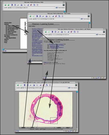
The following figure (Figure #1) is provided to illustrate how the user can move from the introductory pages of the database, into
an image page, and then back out again.

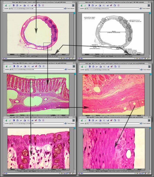
The following figure (Figure #2) is provided to illustrate how the user can move from one image page to another. For any image, the user can switch between unlabeled and labeled versions.
For any image that has a hotspot defined, the user can click in the area to move to an image page of higher magnification. To see all the images for a section, the user should explore all possible branches for each slide in each collection.

A general list of all main pages (including appendix material) in this database is available from the Site Map.
Go to:
[Front page||Main Index|Anatomical Figure|Schematic/Chart|Thumbnail|Slide Number|Keyword]
© 2002 Oxford University Press


 ]. White arrows link to light microscopy images, while black arrows link to electron microscopy images.

