![]()
Departments


![]()

|
27 August 1999 |
Vol. III, No. 4 |
Feature
Article
Science team probing the brain connection
By David J. Craig
|
|
|
A three-dimensional
reconstruction of a neuronal dendrite, whose
branches, called dendritic spines, receive nerve
impulses.
|

Kristen Harris, CAS professor of biology, Malvin Teich, ENG professor of electrical and computer engineering, and Bahaa Saleh, ENG professor and chairman of the electrical and computer engineering department, will use a $960,000 grant over the next five years to develop a revolutionary new method of microscopy and attempt to unravel complex issues in neuroscience.
The scientists will apply advanced theories in quantum physics to observe what occurs in brain synapses -- the sites of communication between neurons. These observations will add to the knowledge of brain function, with particularly important implications for understanding mental retardation.
The grant is funded by the David and Lucile Packard Foundation, through the Interdisciplinary Science Program, which was created in 1998 to help scientists from different disciplines jointly pursue complicated research problems. BU was one of 11 universities and research institutions to receive a grant.
|
|
|
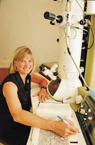
CAS Biology Professor Kristen
Harris studies the finer points of brain function
in a laboratory at Children's Hospital in Boston.
Photo by Kalman Zabarsky
|
"We know these spines are crucial to cognitive processes such as learning and memory," says Harris, "but we know little about how they work, in part because they are so small that they challenge existing optical-imaging techniques."
Harris hopes that her research will produce valuable information about the healthy development of dendritic spines, which when malformed are implicated in causing mental retardation. Little is known about how to ensure the healthy development of the spines.
"A lot of studies are being conducted to find out what molecular biological cues are needed to change spines from their early morphology," she says.
The new imaging technique that the researchers will use to observe the spines is called entangled-photon microscopy, for which Teich and Saleh own a patent. It uses an unusual light source -- a weak beam of photon pairs generated by laser light passed through a nonlinear optical crystal. The photon pairs are expected to provide better resolution than traditional microscopes and reduce light damage to specimens.
|
|
|
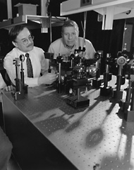
Bahaa Saleh (left), chairman of
ENG's department of electrical and computer
engineering, and Malvin Teich, ENG professor of
electrical and computer engineering, in the Quantum
Imaging Lab at the BU Photonics Center.
BU Photo Services
|
At present, says Harris, specimens cannot remain beneath a microscope for long before the light damages the tissue.
"With an electron microscope, you can see into the individual synapses and cells, but the tissue is dead and its physiological structure is fixed at a certain time point," she says. "But when we look at living tissue, what we see is its gross contours. Now, with this collaboration, we're in a position to use this new way of imaging, which we hope will allow us to visualize the living cell in its native state."
Saleh says that their collaborative effort at BU is unique because they are ideally suited to join advanced physics concepts with imaging technology. "We are a bit ahead of some other institutions," he explains, "thanks to our long-standing interest in both the fundamental quantum physics of light and the optical engineering of imaging systems. That is precisely the mandate of our Quantum Imaging Laboratory." He expects that the group's collaboration will lead to the development of a new instrument that will, in fact, find use in many disciplines within the physical as well as biological sciences.
Harris thinks that such a tool would have profound consequences in biological research, where imaging technology is becoming increasingly important.
"For years, anatomical work was out of favor because much of it was descriptive and didn't tell us enough about function," she says. "But now, with the explosion of molecular approaches, scientists are recognizing that our knowledge of even the basic structure of synapses and connectivity in the brain is quite limited. So all of a sudden scientists who weren't doing anatomy are into imaging."
And Harris, who is relocating her research team from Harvard to BU this fall, anticipates that her research with Teich, Saleh, and Sergienko will lead to further collaborations with researchers in other fields as well. She says that the imaging technique being developed to study dendritic spines might eventually be used to observe cells in a living animal.
"Ultimately, we want to know how the brain works in a living animal, not just how a slice of brain works," she says, "and to link that to behavioral consequences."