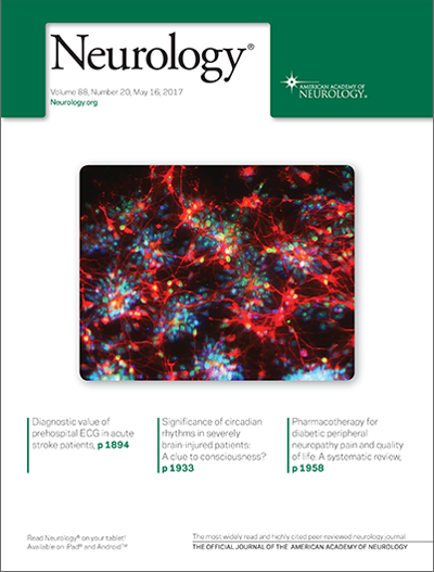
Establishing Gulf War Illness Human Stem Cell Biorepository.
 A team that includes School of Public Health researchers has created a biorepository of stem cells from the blood of veterans with Gulf War Illness (GWI) that will be shared with other researchers and used for mechanistic studies and to test therapeutic compounds, according to a new paper in the journal Neurology.
The team, which includes Kimberly Sullivan, research assistant professor of environmental health and principal investigator on the multi-site Gulf War Illness Consortium, is obtaining peripheral blood cells from Gulf War veterans with GWI and those without, as controls, and turning them into human-induced pluripotent stem cells (hiPSCs), in collaboration with the BU CReM center.
It is the first time that human-induced neurons will be used to try to identify alterations in axonal transport, microtubule functioning and neuroinflamnati0n that may contribute to deficits in cognitive functioning and other symptoms of GWI. This type of study has not been possible due to a lack of available veterans’ post-mortem brain tissue.
Sullivan said the biorepository “will now allow researchers to use both human and animal neurons as a cross-validation of results in translational biomarker studies.” In addition, she said, human-induced neurons are a better path toward developing and testing therapies, given that some features of human neurodegeneration are not reflected in animal models.
The initial hiPSC studies will be conducted at Drexel University and will then be made available to other interested researchers.
The US Department of Defense has funded the research team to recruit blood donors as a follow-up study in a subgroup of 300 participants in the GWI consortium, which also has a biorepository of stored blood, brain imaging and demographic information. While the researchers acknowledge that the number of cell lines they are funded to study is not large enough to evaluate complex factors such as race, gender or genetic background, they say clinical data obtained from the veterans, such as from brain imaging, may yield some “initial insights int0 the severity and nature of the disease corresponding to each of the cell lines.”
In additi0n, they said, “there may be something to be learned from comparing hiPSC cells from equally exposed veterans who either did or did not get sick, given that genetic and possibly epigenetic factors potentially relevant to disease susceptibility would be preserved.”
Recent clinical studies of brain imaging and cognitive functioning have shown significant evidence of altered central nervous system functioning in veterans with GWI sympt0ms. While rodent models have been useful in identifying the effects of particular exposures, the researchers said they are optimistic that using human cell types, such as neurons, glia and muscle, will allow for more opportunities to explore and explain the associations between toxicant exposures and health problems in the GW veterans themselves.
GWI afflicts at least one in four veterans who served in the 1990-1991 war in the Persian Gulf and has been linked to exposure to pesticides, anti-nerve gas pills, and low-level nerve agents, including sarin/cyclosarin. It is a multi-symptom disorder characterized by fatigue, joint pain, and cognitive and gastrointestinal problems, among other symptoms.
Co-authors on the paper, which is on the front cover of the May issue of Neurology, are from Drexel University. The corresponding author is Peter Baas, professor and director of the graduate program in neuroscience at Drexel.
Comments & Discussion
Boston University moderates comments to facilitate an informed, substantive, civil conversation. Abusive, profane, self-promotional, misleading, incoherent or off-topic comments will be rejected. Moderators are staffed during regular business hours (EST) and can only accept comments written in English. Statistics or facts must include a citation or a link to the citation.