
Viscoelastic
| Stabilize the syringe | |
| Inject viscoelastic via syringe |
|
|
| Injection of viscoelastic, notice small air bubble which is removed by a wave of viscoelastic advancing towards the paracentesis. |
The injection of viscoelastic may also seem trivial, but a few key reminders can prevent avoidable complications.
Instrumentation

Figure 1: A dispersive viscoelastic coats the endothelium well, but “disperses” making removal more difficult.
Choose your viscoelastic wisely. At this step, we typically use Viscoat (Alcon Laboratories, Inc, Fort Worth, Texas), a dispersive viscoelastic that coats the endothelium and provides endothelial protection during phacoemulsification (Figure 1). Later in the surgery prior to lens insertion, we typically prefer a cohesive viscoelastic, such as Provisc (Alcon Laboratories, Inc, Fort Worth, Texas). Cohesive viscoelastics maintain the chamber yet are removed easily at the end of the case (Figure 2).
There are a number of different approaches to this step, including a “soft shell” technique combining the endothelial coating properties of a dispersive viscoelastic with the improved capsular handling properties of a cohesive viscoelastic, or newer viscoelastic agents exhibiting both cohesive and dispersive properties. Most agents are distributed with a syringe and cannula, which can be easily assembled prior to use.
Technique
Prior to entering the eye, ensure the viscoelastic flows smoothly from the syringe while also removing all air from the syringe and cannula. Simply injecting a small amount of viscoelastic at the limbus can accomplish this task, and will help prevent the introduction of air bubbles into the anterior chamber. Bubbles primarily will affect the view during capsulorrhexis, and thus should be avoided.
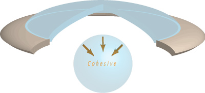
Figure 2: A cohesive viscoelastic maintains space and thus the anterior chamber, and “sticks together” thus easing removal.
Any pressure on the posterior aspect of the wound will cause efflux of aqueous, and therefore care must be taken not to deflate the anterior chamber. This can be accomplished by maintaining the cannula within the wound and avoiding downward pressure at the wound site. Also, while entering the eye, begin injecting viscoelastic immediately to provide an advancing “shield” of viscoelastic, to prevent inadvertent contact with the lens capsule or endothelium.
Once the eye is entered, continue injecting while advancing the tip all the way across the anterior chamber. More forceful injection can now proceed, while withdrawing the cannula. This technique ensures adequate filling of the chamber, while also ensuring that the advancing wave of viscoelastic propels remaining aqueous and air towards the paracentesis and out of the eye.