Announcing the 2017 MSE Innovation Grant Winners
Congratulations to the winners of the 2017 MSE Innovation Grants!
The idea behind BU MSE Innovation Grants Program is to create a low overhead way of encouraging innovation and risk taking in a national funding environment that makes this increasingly difficult. Each year we will be awarding about five awards of approximately $10k each. Any faculty member (any rank) in the extended BU MSE community is eligible for the award. It is a one time, non-recurring award that can be used for equipment, salary for a student or post doc, travel by a BU person to another location, travel by someone else to BU or any other legitimate research expense. The idea is to enable real innovations to take place and encourage far out thinking.
2017 MSE Innovation Grant winners:
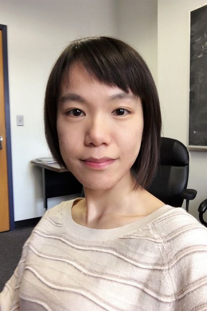
 Xi Ling (Chemistry, MSE)
Chemical Vapor Deposition of Two-dimensional Boron Monolayer and Heterostructures
Two-dimensional (2D) boron joined the family of 2D materials recently, and attracted much attention in a short time. There are many boron polymorphs with different properties from metal to semiconductor. Among them, g phase 2D boron monolayer shows semiconducting properties with direct band gap at about 2.25 eV, which indicates that it has giant potential in optoelectronic devices. Also, the stability in air and anisotropic structures make it more applicable for multifunctional devices. However, obtaining the high quality and large-area 2D boron monolayer is still in the early stage. Here, I propose to use multi-zone chemical vapor deposition method to synthesize the 2D boron monolayer crystal and potentially hybrid it with other 2D materials developing in our lab to form the heterostructures. The successful growth of the 2D boron will open the door for the properties and applications exploration, and facilitate the collaboration with other colleagues at BU in the future.
 Rama Bansil (Physics, MSE)
Microfluidic Studies of Bacteria Chemotaxis in Viscoelastic Polymer Solutions and Gels
Many bacteria move through highly viscous environments to reach their colonization niche. In particular, we focus on the gastric ulcer and cancer causing bacterium Helicobacter pylori, which moves across the viscolealstic, gel-like mucus layer in the stomach to colonize on the epithelial cell surface lining the stomach wall. We have examined the swimming behavior of these helical-shaped bacteria by using video optical microscopic to track them in various solutions and gels. The innovation project goes a step further by using microfluidic approaches to mimic the physiological situation where the bacterium directs its motion from the cavity of the stomach towards the epithelial cell surface, using gradients in concentration of chemicals such as urea and gastric juice secreted by the stomach glands. These studies will help us understand not only how bacteria swim in response to chemical gradients, but also how they in turn influence the rheological properties of their host medium to create a unique, locally structured environment best suited for their own survival.
 Chuanhua Duan (ME, MSE)
Rapid and Scalable Patterning of Conjugated Polymer Thin Films With Controlled Morphology
Conjugated polymers (CPs) are organic macromolecules that are characterized by a backbone chain of alternating double- and single- bonds. Because of their high solution processability and excellent semiconductor properties, CPs, especially in their thin film forms, have found various promising applications in electronics, photonics and photovoltaics. Further development of CB-based technologies requires addressing two critical manufacturing challenges of CP thin films: 1) accurate control of CP thin film morphology including molecular conformation and inter-chain structure; 2) rapid formation of patterned CP thin films with high spatial resolution and controlled thickness. Since these two challenges cannot be simultaneously resolved by existing manufacturing methods, we propose to prepare patterned CP thin films using a new technique titled pervaporation-assisted molding in nanofluidic channels. In this technique, polymer solution will be introduced into nanochannel molds that are temporarily bonded to the device substrates. Pervaporation will be employed on the sidewall of the nanochannels to rapidly remove solvent and promote thin film self-assembly with controlled morphology along the nanochannels. We will systematically investigate the speed, morphology control and scalability of this new technique. Success of this proposed research would result in an ideal manufacturing technique for thin film formation and patterning, which will not only boost CP-based organic electronics and photonics, but will also advance other emerging applications involved with patterned polymer/nanoparticle thin films.
 Douglas Holmes (ME, MSE)
Morphing and Growth of Soft Structures
Soft and thin structures can dramatically deform in response to small amounts of external force, and controlling these deformations has been the focus of significant research efforts among physicists, biologists, and engineers in the last decade. The ability to produce a generic structure that can be morphed into a desired shape has important implications for a wide array of industries, ranging from hierarchical manufacturing to deployable structures and actively morphing wings. By tailoring the swelling and growth of soft materials, we aim to create deformable structures that can be reconfigured into a desired shape by altering their local geometry with various stimuli, e.g. chemical, electrical, thermal. We have demonstrated the controlled morphing of sheets into shells by using a novel approach of residual swelling, where rubber plates are prepared with a localized excess of free monomer chains. The diffusion of this residual solvent locally stretches and shrinks the sheet causing it to morph into a 3D shape. The resulting structure is a geometric composite – it combines different intrinsic geometries within a material to produce shapes that differ from their individual components. With this research endeavor, we will study and quantify the connection between geometry and the resulting structural and dynamical instabilities of geometric composites. This mechanistic understanding is necessary to develop technological pathways for advanced, active structures that operate in complex environments, and will provide fundamental insights into the connection of geometry and topology for morphing and design of multifunctional soft materials.

 Alice White (ME, MSE) and Ren Zhang (ME)
Dynamic 3D Scaffolds for Tissue Engineering
In vivo, cells are in a dynamic 3D environment with biochemical and physical cues. The physical properties (i.e., mechanical stiffness, topography) of the extracellular matrix play an important role for cell migration, proliferation and differentiation. However, it is challenging to create a cell culture environment with tunable mechanical properties and dynamic actuation capability for more physiologically relevant in vitro model development. We propose to fabricate cell scaffolds through direct laser writing (DLW) based on two-photon polymerization (TPP) in flexible hydrogels, which will be subsequently coated with a thin biocompatible metal oxide layer using atomic layer deposition (ALD). In this way, the structure design flexibility with submicron resolution and the potential piezoelectric actuation properties can be combined to generate an active and responsive cell culture environment. The dynamically movable in vitro 3D structures would mimic the real cell surroundings in tissues, and thus facilitate the development of biocompatible nano-platforms for clinical translation.

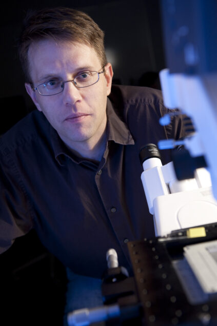 Michael Albro (ME, MSE) and Sean Andersson (ME, SE)
Fluorescent Colocalization Microscopy for Novel Quantification of Cellular Activation of TGF–β