Laboratory Testing
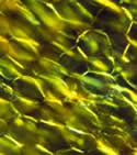 The sine qua non for the diagnosis of amyloidosis is a tissue biopsy staining positive with Congo red and demonstrating green birefringence under polarized light microscopy. Electron microscopy will also show a classical fibrillar appearance in the extracellular matrix.
It is important to avoid over-staining the tissue with Congo red as this may give false results. The technique of subcutaneous fat pad aspiration offers a simple and relatively non-invasive approach to the diagnosis of systemic amyloidosis and is positive in 70-80 % of patients with systemic amyloidosis. If the fat is negative, but suspicion of disease is high, a biopsy of an involved organ system (e.g., heart, kidney, or liver) may need to be done to make the diagnosis. An involved organ is positive nearly 100% of the time.
Before a patient is evaluated at the Amyloidosis Center, we request of review of the slides and, wherever possible, an unstained block of tissue so that a standardized appropriate stain can be done. Tissues and slides will be returned after evaluation. We ask that you call to discuss testing requests before sending samples.
Address for sending biopsy material for a second opinion which should always be accompanied by an Amyloid Pathology Consultation Form:
BU Pathology
670 Albany Street
Boston, MA 02118
Phone: (617) 414-7089
Sending Tissue Samples for Amyloid Histopathology
Abdominal Fat Aspirate Procedure
Abdominal Fat Aspirate Staining Technique